Research Article | Volume: 12, Issue: 5, May, 2022

Quillaja saponaria bark saponin attenuates methotrexate induced hepatic oxidative stress, inflammation and associated liver injury in rats

Mustafa Ahmed Abdel-Reheim Ahmed Amine Ashour Mohamed Abdelrazik Khattab Ahmed Gaafar A. Gaafar   

Open Access   

Published:  May 05, 2022

DOI: 10.7324/JAPS.2022.120510
Abstract

Quillaja saponaria Molina tree bark represents the main source of Q. saponaria saponin that has antitussive-, antiinflammatory-, and immune-stimulant effects. In spite of such multiple applications, the potential role of Q. saponaria bark saponin (QBS) as a hepatoprotective agent has not been elucidated. On the other hand, methotrexate (MTX) is an antineoplastic/immunosuppressive drug that may cause serious liver complications which limit its clinical use as a result of the arising oxidative stress, inflammation, and apoptosis. This study aimed to investigate the protective effects of QBS against MTX-induced hepatotoxicity. Thirty-two male rats were divided into four groups (N = 8). Control rats received oral saline (0.2 ml/day, for 10 days) (group I). In group II, rats were administered oral saponin (100 mg/kg/day, for 10 days). In group III, rats received saline (0.2 ml/day, p.o., for 10 days) and then were injected with single i.p. MTX (20 mg/kg) on day 5. Rats in group IV (MTX + QBS) received saponin (100 mg/kg/day, p.o., for 10 days) with single i.p. MTX (20 mg/kg) on day 5. On day 11, blood and livers were collected. Serum alanine aminotransferase (ALT), serum aspartate aminotransferase (AST), serum alkaline phosphatase (ALP), and serum lactate dehydrogenase (LDH) levels were measured as liver-damage biomarkers. Hepatic malondialdehyde (MDA), reduced glutathione (GSH), and total nitric oxide content (NOx ) were determined as oxidative-stress measures. Hepatic Bcl-2 and Bcl-2 associated-X-proapoptotic protein (Bax) mRNAs were assessed as apoptosis biomarkers. Hepatic nuclear factor erythroid 2-related factor-(Nrf-2)/heme oxygenase-1 (HO-1) immunoreactivities were evaluated as indicators for inflammation. Interestingly, QBS attenuated MTX-mediated elevations of AST, ALT, ALP, LDH, MDA, NOx , and Bax, while inhibiting MTX-induced decreases in GSH, Nrf-2, HO-1, and Bcl-2. Histological analysis further confirmed the hepatoprotective microenvironment rendered by QBS. Conclusively, our findings represent the first evidence that QBS might confer valuable hepatoprotection against MTX-mediated liver damage through suppression of oxidative stress, apoptosis, and amelioration of tissue inflammation, proposing QBS as an effective therapeutic regimen to be involved in future hepatic-support therapies.


Keyword:     Quillaja saponaria methotrexate hepatotoxicity oxidative stress apoptosis Nrf-2/HO-1


Citation:

Abdel-Reheim MA, Ashour AA, Khattab MA, Gaafar AGA. Quillaja saponaria bark saponin attenuates methotrexate?induced hepatic oxidative stress, inflammation and associated liver injury in rats. J Appl Pharm Sci. 2022; 12(05):129–141.

Copyright: © The Author(s). This is an open-access article distributed under the Creative Commons Attribution License, which permits unrestricted use, distribution, and reproduction in any medium, provided the original work is properly cited.

HTML Full Text

INTRODUCTION

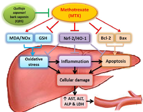
Drug-mediated adverse effects and associated toxicities are some of the most frequently experienced problems during medication practice. Several drugs have great therapeutic advantages in the management of several disorders, but unfortunately, their pharmacological use is notably limited by their correlated adverse reactions (Gamal Helal and Said, 2020). Methotrexate (MTX) is an example of those drugs which is used as a chemotherapeutic agent for the treatment of cancer and as an immunosuppressant agent in the treatment of rheumatoid arthritis, ectopic pregnancies, and other diseases (Malaviya, 2016) MTX -mediated anticancer and immunosuppressive effects are owing to its function as an antagonist for folic acid (competitively inhibits dihydrofolate reductase, the enzyme that catalyzes the conversion of dihydrofolate to tetrahydrofolate), which in turn prevents cellular mitosis by inhibiting thymidylate, purines, and folic acid required for DNA synthesis (Gibson et al., 2019), preventing cells from dividing (Liu et al., 2019). However, acute MTX-associated vital organ toxicity, including kidney, liver, gastrointestinal mucosa, and bone marrow (Bhatnagar et al., 2015; Fridlington et al., 2011), is the rate-limiting factor for its clinical use (Malaviya, 2016). Decreasing MTX toxicities and, in turn, restricting the discontinuation of MTX therapy is a current therapeutic challenge.

Importantly, hepatotoxicity is a serious intricacy of chronic MTX therapy, which is accompanied by the elevation of liver enzymes (Sotoudehmanesh et al., 2010). Although the main mechanism(s) of MTX-mediated hepatic injury has not yet been fully elucidated, there are previous studies which hypothesized that the arising oxidative stress gives rise to the formation of a copious amount of free radicals with concomitant lipid peroxidation of biological membranes as one of the main reasons (Ekinci-Akdemir et al., 2018). Emerging evidence suggests that such oxygen-derived free radicals substantially contributed to the etiology of liver injury and other types of tissues’ destruction (Oyagbemi et al., 2016). Indeed, MTX induces oxidative stress via two pathways: first, excessive production of reactive oxygen species (ROS) through dysfunction of mitochondria and respiratory chain defects (Famurewa et al., 2017); second, through the depletion of reduced glutathione (GSH) in the attribution of inhibition of reduced nicotinamide adenine dinucleotide phosphate production (Al Maruf et al., 2018; Salimi et al., 2019). GSH is an important nonenzymatic antioxidant bioactive peptide that appears in the liver, which binds toxic substances, such as electrophilic radicals and ROS, originating from extensive oxidative effects (Chen et al., 2013). Nitric oxide (NO) is another highly interactive pro-oxidant produced by liver parenchymal and nonparenchymal cells which acts as an important mediator of several physiological and pathological events. The reactivity of NO with ROS indicates many biological cascades through which NO might either enhance or decrease oxidative stress-induced tissue injury (Ozcelik et al., 2014). The oxidative inflammatory consequence is a common feature of chronic diseases. During inflammation, the immune system cells are recruited at the injury site leading to sudden increase of cellular respiration and oxygen consumption, thus accumulating ROS at the injury site (Reuter et al., 2010). Previous studies proved that Nrf-2 contributes to the anti-inflammatory process by regulating gene expression through the antioxidant responsive elements (ARE) including heme oxygenase-1 (HO-1) and orchestrating the recruitment of inflammatory cells (Tu et al., 2019). Such inducible HO-1 catalyzes the degradation of heme (a cellular pro-oxidant toxicant) to biliverdin, carbon monoxide, and free iron. Biliverdin is subsequently metabolized to the antioxidant, bilirubin, that scavenges peroxy-radicals and attenuates lipid peroxidation (Ryter et al., 2002). In addition, HO-1 negatively regulates the inflammatory mediators, interleukin- (IL-) 6 Nuclear factor kappa B (NF-κB), and TNF-α (Aleksunes et al., 2010), as one of the proinflammatory cytokines which is increased after MTX treatment which causes neutrophil infiltration and strikes apoptotic cell death (Cure et al., 2015), in response to such oxidative stress and inflammation (Wang, 2015), with the regulation of the antiapoptotic Bcl-2 family proteins that prevent apoptosis by inhibiting the activity of proapoptotic Bcl-2 associated-X-proapoptotic protein (Bax) protein (Barclay et al., 2015).

In animals, saponins can antagonize the action of the inflammatory mediators (Sagesaka et al., 1996) and possess anticarcinogenic properties (Xu et al., 2016) due to their amphiphilic nature, which causes the destruction of cancer cells (Top et al., 2017). The bark derived from the South American soap tree, Quillaja saponaria Molina, represents the main source of Q. saponaria saponins that have been studied for more than four decades for their relevant biological activities with a wide range of applications. In traditional medicine, the Q. saponaria tree has been used orally to relieve cough/bronchitis, as well as topically to eliminate dandruff and to relieve scalp itching (Leung, 1980). The raw materials have an anti-inflammatory, hypocholesterolemic effect (Fleck et al., 2019), with strong immunostimulant activity that makes it an ideal vaccine adjuvant (Rajput et al., 2007). Historically, the powder of the inner bark has been used as a detergent (owing to the presence of a glycoside saponin) (Fleck et al., 2019). The saponin of the Quillaja bark is a triterpenoid saponin glycoside comprising a hydrophilic sugar moiety and hydrophobic aglycone backbone (sapogenin). The main sapogenin that is most commonly reported is a triterpene termed quillaic acid (Guo and Kenne, 2000; Guo et al., 1998; Osbourn et al., 2011). The Q. saponaria bark saponin (QBS) is structurally distinct from other triterpenoid saponins by exhibiting a fatty acid and an aldehyde group at C4 of the triterpene (Kensil et al., 1995). Although most known saponins cause hemolysis of blood cells (Top et al., 2017), the triterpenoid QBS was reported, interestingly, to be relatively nonhemolytic, with immunostimulant and antioxidant effects (Ahmed Abdel-Reheim et al., 2017). In addition, QBS has been demonstrated to have in vitro antimicrobial (Hassan et al., 2010) and anticancer (Hassan et al., 2013; Hu et al., 2010) activities. Moreover, quillaic acid demonstrated powerful topical in vivo anti-inflammatory efficacy in different mouse models (Rodríguez-Díaz et al., 2011).

The MTX-induced hepatic, hematologic, and immune complications along with the QBS-mediated anti-inflammatory-, antioxidant-, and immune-stimulant effects are observations which attract our attention to the possibility of using QBS as an adjuvant therapy with MTX. In fact, no studies, to date, have investigated the role of Q. saponaria bark extracts in alleviating MTX-mediated liver injury. Accordingly, we tried to build a rationale for using such saponin to counteract the deleterious effects of MTX, and the current study was designed to investigate the virtual hepatoprotective activity of the saponin derived from the Q. saponaria bark against MTX-mediated liver injury and to highlight some of the possible molecular mechanisms underlying these effects. To fulfill such aims, biochemical markers illustrating the loss of liver integrity, including serum aspartate aminotransferase (AST), Serum alanine aminotransferase (ALT), serum lactate dehydrogenase (LDH), and serum alkaline phosphatase (ALP), were assessed, and tissue redox biomarkers, MDA, GSH, and total nitric oxide (NOx) content, were determined. Furthermore, the levels of hepatic Bcl-2 and Bax mRNA expression were assessed for determining the level of apoptosis, and histological studies were executed to assess the extent of histopathological lesions associated with adopted treatments. Finally, histochemical immunoreactivity of Nrf-2/HO-1 proteins was utilized to validate the involvement of the inflammatory responses of the designated treatments.


MATERIALS AND METHODS

Ethics statement

All procedures in the current study were approved by the Research Ethics Committee of the Experimental Animals Use and Care, Faculty of Pharmacy, Beni-Suef University (REC-A-PhBSU). The experimental protocol of laboratory animals handling was in accordance with the approved guidelines of the Animal House Rules of the Dept. of Pharmacology and Toxicology, Section B, Faculty of Pharmacy, Beni-Suef University in 2009, that follows the conventional guidelines of the National Institutes of Health (NIH), Eighth Edition, The National Academies Press, Washington, DC, revised in 1985, for the care and use of laboratory animals.

Animals

Male Wistar rats (weight, 150–180 g; age, 6–8 weeks) were obtained from the National Research Center, Cairo, Egypt. The rats were kept in plastic cages and maintained in an animal care facility under standard conditions of humidity, temperature (25°C ± 1°C), and 12 hours light/dark cycles. Rats were allowed free food/water access and were left for 7 days before the treatment to acclimatize.

Drugs

Methotrexate

MTX was obtained from Baxter Company (Cairo, Egypt) and was singly injected into the animals (i.p., at a dose of 20 mg/kg on the fifth day) (Fouad et al., 2020).

Saponin from Q. saponaria Molina

QBS powder was purchased from Sigma-Aldrich (St. Louis, MO). After dissolving it in normal saline, the resultant solution was given orally to the animals (at a dose of 100 mg/kg/day, for 10 days) (Ahmed Abdel-Reheim et al., 2017). The main aglycone (sapogenin) that is most commonly reported in QBS is the triterpenoid, quillaic acid, a pentacyclic triterpene, a hydroxy monocarboxylic acid, and an aldehyde of predominantly 30-carbon atoms of the Δ12-oleanane type (C30H46O5), that is characterized chemically and using nuclear magnetic resonance spectroscopy and mass spectrometric methods (Guo et al., 1998). QBS is soluble in water with an average MW of 56,000. QBS solutions are stable for about 1 month when stored at 2°C–8°C, while QBS powder is stable for at least 1 year at room temperature when stored dry (Sigma Product No. S4521).

Preparation of the sapogenin

According to SIGMA Product Specifications, sapogenin devoid of any sugars is isolated from the bark of the South American soap tree, Q. saponaria Molina (Rosaceae family), by acid hydrolysis of saponins, and purified by ultrafiltration to reduce low-molecular-weight contaminants (Dalsgaard, 1978). The sapogenin content is ≈35%. The isolation, quantification, and quality control of the purified acylated triterpenoid saponin were reported in the Product Information Data Sheet (Sigma Product No. S4521).

Chemicals and kits

AST and ALT reagents kits were purchased from Diamond Diagnostics, Cairo, Egypt. ALP kits were obtained from Biodiagnostics, Cairo, Egypt. LDH kits were obtained from Biosystems (Barcelona, Spain). N-(1-Naphthyl) ethylenediamine dihydrochloride, Ellman’s reagent, MDA, reduced GSH, and thiobarbituric acid were obtained from Sigma-Aldrich (St. Louis, MO). 3,3?-Diaminobenzidine tetrahydrochloride (DAB) was obtained from Vector Laboratories Inc., Burlingame, CA. Nrf-2 and HO-1 rabbit polyclonal and monoclonal antibodies, respectively, were obtained from Abcam Biochemicals (Cambridge, MA). The GF-1 total RNA extraction kit was obtained from Vivantis Technologies Sdn Bhd (Malaysia). The complementary DNA (cDNA) synthesis kit was obtained from Bio-Rad, CA. All other used chemicals were of the highest standard analytical grade.

Experimental design

Thirty two rats were randomized into four groups, eight rats in each. Group I served as control rats and received oral 0.9% normal saline (vehicle of saponin; 0.2 ml/day, for 10 consecutive days), in addition to a single i.p. injection of 0.9% saline (vehicle of MTX) on day 5. Group II of rats (saponin alone-treated group, QBS) was administered oral QBS (100 mg/kg/day) dissolved in normal saline for 10 consecutive days, with an additional single i.p. injection of 0.9% saline on day 5 only. Group III of rats was administered oral 0.9% normal saline (for 10 consecutive days), in addition to a single i.p. injection of MTX (20 mg/kg) on the fifth day of the experiment. In group IV (MTX + QBS), the rats received the saponin (100 mg/kg/day, p.o., for 10 consecutive days) along with a single i.p. dose of MTX (20 mg/kg) on the fifth day of the experiment, 2 hours before saponin administration.

Twenty four hours after the last dose of the saponin treatment, animals were anesthetized using thiopental sodium (i.p., 75 mg/kg), and the blood samples were withdrawn using heparinized microcapillary tubes from the retroorbital plexus, collected in centrifuge tubes, left at room temperature to coagulate and put in a water bath at 37°C for 10 minutes and then centrifuged at 1,000 g for 20 minutes. After centrifugation, the separated serums were collected and stored at −20°C until the assessment of AST, ALT, ALP, and LDH levels (Kiran et al., 2012). After blood assembling, rats were sacrificed by cervical dislocation, and the livers were immediately dissected out and washed three times with ice-cooled normal saline (Kiran et al., 2012). Each liver was cut up into three sections. One section was fixed in 10% phosphate-buffered formalin for histopathological and immunohistochemical examinations of Nrf-2/HO-1. The second part was homogenized (1/5 w/v) in ice-cold Tris-HCl buffer (0.1 M, pH 7.4) for preparation of 20% tissue homogenates and was kept at −20°C for biochemical assays. The third part was shock-frozen in liquid nitrogen and was kept at −80°C for quantitative real-time-polymerase chain reaction (qRT-PCR) analysis for determination of Bcl-2 and Bax mRNA expression levels. This model was adopted after Mehrzadi et al. (2018) with slight modifications.

Biochemical assays

Serum biomarkers assessments

Serum biomarkers were determined using commercial kits; serum AST and serum ALT were determined according to the Reitman and Frankel (1957) method; serum ALP was determined according to the Belfield and Goldberg (1971) method; serum LDH was determined according to the Vassault (1983) method.

Oxidative stress parameters assessments

To prepare a 20% tissue homogenate, 1 g of each liver was homogenized with five volumes of ice-cooled phosphate-buffered saline (PBS) using an IKA homogenizer (Model T 25 ULTRA-TURRAX, Staufen, Germany). About 2 ml from each of the obtained homogenates was moved into a 2 ml Eppendorf tube and centrifuged (at 700× g), and the obtained supernatants were used for the determination of hepatic GSH, according to the Sedlak and Lindsay (1968) method; hepatic MDA, according to the Uchiyama and Mihara (1978) method; and NO content, according to the Miranda et al.’s (2001) method.

Histological investigations

Histopathological analysis

The isolated portions of the liver were washed using normal saline, kept in 10% phosphate-buffered formalin for 72 hours in well-sealed containers, dehydrated with a sequence of graded alcohol solutions, cleared in xylene, and infiltrated by Paraplast tissue embedding media (Sigma-Aldrich, St. Louis, MO). Paraffin-embedded tissue blocks were sectioned (using a sledge microtome), and 4 μ thick sections were put on glass slides, deparaffinized, stained with hematoxylin and eosin dye, and coded for blind assessment of microscopic analysis of liver histology under a light microscope (Leica Microsystems GmbH, Wetzlar, Germany). The liver injury was assessed by a hepatopathologist who was unaware of the treatments that had been received. For semiquantitative evaluation of common indices (e.g., degenerative hepatocytes, congestion of hepatic sinusoids/vasculature, inflammatory cell infiltration, and cellular apoptosis/necrosis), the scoring system was adopted as follows: 0> normal, 1> mild, 2> moderate, and 3> severe, and the total score was determined by averaging all scores of each group (Michael, 2008). The microphotomicrographs (×100 magnification) were encoded on 24 bits/pixel on three channels (blue, green, and red), and the representative-colored images were shown. All standard procedures for samples fixation and staining were according to Culling (1974).

Immunohistochemical determination of Nrf-2 and HO-1

Immunohistochemical staining was conducted as follows: Paraffin-embedded 4 μm thickness tissue sections of different groups were deparaffinized and rehydrated. The endogenous activity of peroxidase was occluded by adding 3% H2O2 for 20 minutes, followed by a 15 minutes wash in PBS. Antigens were retrieved by placing the sections in a 10 mM sodium citrate buffer (pH 6.0) and warmed up at 121°C in a hot oven for 30 minutes. Then, sections were cooled at room temperature for 20 minutes and rinsed in PBS. For blocking nonspecific protein binding, sections were incubated for 1 hour in Tris-buffered saline (containing 5% bovine serum albumin). Sections were then incubated overnight (at 4°C) with the primary rabbit polyclonal antibody against rat Nrf-2 from Abcam Co. (ab92946) or rabbit monoclonal antibody against rat HO-1 from Abcam Co. (ab13248) (dilution 1:2,000). The slides were then washed by PBS before incubation with the secondary antibody, HRP Envision Kit (DAKO), for 20 minutes. The slides were then washed with PBS. The positive immunoreactivity was developed and visualized with 0.05% DAB color reaction for 15 minutes. After washing with PBS, the sections were counterstained with hematoxylin for staining nuclei, dehydrated, and cleared in xylene. The cover-slipped tissue sections were observed for brown color formation (that was considered as an indicator of protein expression) under a light microscope. The stained slides were coded before examination by a specialist who was blinded to the treatment protocol. Morphological assessments and data analysis were attained using the Leica Application module for tissue sections analysis that is connected to a full HD microscopic imaging computer system (Leica Microsystems GmbH, Germany). Quantitative determinations of protein expression levels of Nrf-2 or HO-1 were done by estimation of mean area percentage of Nrf-2 or HO-1 immunoexpression using a microscopic camera attached to the Leica software that converts the measured units (pixels) given by the image-analyzer program into actual micrometer units used for immunoexpression analysis. Six nonoverlapping fields in the immunostained tissue sections were randomly selected and acquired from each sample for quantification of positive immunoexpression percentage levels of Nrf-2 and HO-1.

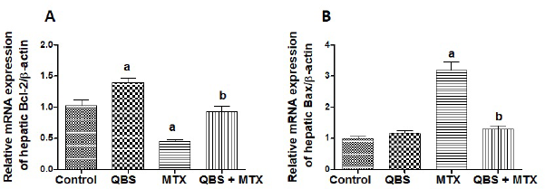
Real-time PCR and determination of hepatic mRNA levels of Bcl-2 and Bax

Total RNA was isolated from hepatic tissues using GF-1 total RNA extraction kit (GF-TR-050, Vivantis Technologies Sdn Bhd, Malaysia) according to the manufacturer’s instructions. To get rid of the contaminating DNA, the isolated RNAs were treated with a DNase I, RNase-free kit (Fermentas, MD). One microgram of the total RNA and random primers were used for cDNA synthesis by adopting the Script™ cDNA synthesis kit (Bio-Rad, CA). For qRT-PCR, the cDNA samples were run in triplicate, and the PCR reactions were carried out using Power SYBR® Green (Life Technologies, CA) and were attached to the Applied Biosystem, version 3.1 software (StepOne™) (CA). The typically adopted thermal profile was 95°C for 5 minutes (for denaturation), followed by 45 amplification cycles of 95°C for 30 seconds (denaturation step), 56°C for 30 seconds (annealing step), and 72°C for 30 seconds (elongation step). After PCR amplification, the values of ΔCt were calculated by subtraction of the β-actin Ct from the Ct of each sample, and the relative levels of gene expression were determined. β-actin is used as a reference gene. The used primers sequences are shown in Table 1.

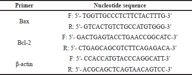
Table 1. The sequences of the primers used in the current study.

[Click here to view]

Statistical analysis

Values were expressed as means ± SEM. Multiple comparisons between different treatment regimens were obtained using the one-way analysis of variance (ANOVA) test followed by the Tukey-Kramer comparison tests that were applied across the four groups or using the chi-squared (χ2) test, wherever indicated. The results were considered significantly different at p < 0.05. Data analysis was accomplished using the computer software GraphPad Prism and GraphPad InStat, San Diego, CA.


RESULTS

Treatment with QBS ameliorated MTX-induced hepatic damage and improved liver integrity profile

Alterations in serum levels of AST, ALT, LDH, and ALP might indicate liver dysfunction. As shown in Table 2 and Figure 1, MTX significantly elevated serum AST, ALT, ALP, and LDH levels in MTX-treated rats as compared to their corresponding levels in the normal rats from the control group. Pretreatment with QBS significantly attenuated MTX-induced elevations in serum levels of AST (by ~43%), ALT (by ~40%), ALP (by ~23%), and LDH (by ~46%), as compared to MTX-treated rats. Furthermore, rats administered QBS did not exhibit change in the levels of the indicated biomarkers when compared to the normal control group of rats. Such results propose the hepatoprotective activities of this saponin against MTX-mediated damage of liver cells.

Table 2. The effects of QBS treatment on the serum levels of AST, ALT, ALP, and LDH in MTX-treated rats.

[Click here to view]

Figure 1. The modulatory effects of QBS pretreatment on the serum levels of AST (A), ALT (B), ALP (C), and LDH (D) in MTX-treated rats. The depicted data describes the fold changes ± SEM of the levels of the demonstrated biomarkers in the serum of the rats from the indicated groups (N = 8). Statistical inspections were accomplished using ANOVA followed by Tukey’s multiple-comparison test; a p < 0.05, versus normal control animals; b p < 0.05, versus MTX-treated animals.

[Click here to view]

Table 3. The effects of QBS on the oxidative stress biomarkers (hepatic contents of GSH, MDA, and NOx) in MTX-treated rats.

[Click here to view]

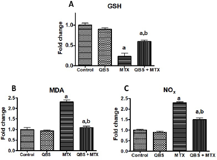
Figure 2. The modulating activities of QBS pretreatment on the oxidative stress biomarkers [hepatic contents of GSH (A), MDA (B), and NOx (C)] in MTX-treated rats. The depicted data describes the fold changes ± SEM of the levels of the demonstrated oxidative stress biomarkers in the hepatic tissues of the rats in the indicated groups (N = 8). Statistical inspections were accomplished using ANOVA followed by Tukey’s multiple-comparison test; a p < 0.05, versus normal control animals; b p < 0.05, versus MTX-treated animals.

[Click here to view]

Treatment with QBS attenuated hepatic oxidative stress and suppressed hepatic lipid peroxidation

GSH is an important nonenzymatic antioxidant that maintains normal cell integrity by binding/inhibiting toxic electrophilic radicals and oxygen free radicals (Chen et al., 2013), most of which have a reactive nature with NO. NO is one of the important oxidant mediators which possesses prominent cytotoxicity and stress-induced cell injury (Ozcelik et al., 2014). Such a chain of oxygen free radicals cascade generates lipid peroxidation in several biological systems with the formation of the final product: malondialdehyde (MDA) (Tsikas, 2017). Hence, decreased hepatic GSH levels with elevated MDA (a measure of lipid peroxidation) and total NOx contents characterize the hepatic oxidative stress (Cruz et al., 2020). With regard to the current study, our data showed that MTX markedly decreased (~77%) liver content of GSH and outstandingly increased hepatic MDA and NOx contents in MTX-treated rats (by about 1.3-fold and 1.4-fold, respectively), in comparison with the untreated rats in the control group (Table 3 and Fig. 2). However, pretreatment with QBS showed a remarkable elevation in the hepatic GSH level (by ~57%) along with a notable decline in both MDA and NOx contents (by ~57% and 36%, respectively) as compared to MTX-treated rats (Table 3). The QBS-mediated improvement of the oxidant/antioxidant profile and the reduction in pro-oxidant load might be accountable for the hepatoprotective effects of the used saponin against liver injury induced by MTX.

QBS mitigated MTX-induced histopathological/inflammatory modifications in the liver tissues

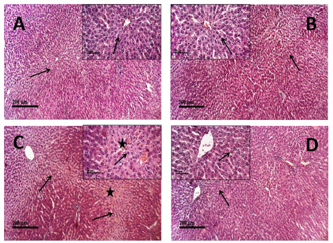
Hematoxylin-eosin (H and E) staining was employed in the current study to assess the pathologic alterations in hepatic tissues dissected from rats exposed to the indicated treatments (Fig. 3A–D). As indicated in Figure 3A, microscopic examinations of liver tissues from vehicle-treated control rats revealed normal histological structures of hepatic parenchyma with intact radiating hepatocytes in hepatic lobules showing large vesicular nuclei with prominent nucleoli (arrow), intact hepatic sinusoids with lining endothelial cells and normal resident Kupffer cells as well as intact vasculatures (score = 0). Rats treated with QBS alone (Fig. 3B) exhibited similar normal histological features of the hepatic lobule with intact hepatocytes (arrow) without abnormal morphological or structural changes (score = 0). Conversely, the liver tissues obtained from MTX-treated rats showed moderate centrilobular vacuolar degenerative changes (score = 2) of most of the hepatocytes (arrow) in the examined hepatic sections accompanied with obvious congested hepatic sinusoids and central veins (star) revealing sinusoidal leukocytosis with inflammatory cell infiltration (score = 3), along with sporadic hepatocyte apoptosis or necrosis (score = 2) (Fig. 3C). However, pretreatment with QBS extremely mitigated the severity of MTX-produced hepatic pathological conversions. The hepatic sections from rats treated with QBS/MTX showed better organized morphological architecture of hepatic lobules with significant reduction in the congested sinusoids and vasculature (score = 1), a few inflammatory cells infiltration, and only mild sporadic vacuolar degenerative changes (arrow) of hepatocytes (score = 1) (Fig. 3D). Taken together, such observations indicate that QBS treatment dramatically impaired MTX-induced liver injury in the examined rats.

Effects of QBS treatment on Nrf-2/HO-1 pathway in the livers of MTX-treated rats

Nrf-2/HO-1 is a defensive pathway whose activation is increased in various pathological conditions (Aleksunes et al., 2010) to counteract the inflammatory and apoptotic actions of the causative insults (Goodman et al., 2007). In the present work, we hypothesized that Nrf-2/HO-1 contributes in mediating the protective effect of QBS against MTX. To validate the role of the Nrf-2/HO-1 pathway, we further determined the expression of Nrf-2 and its target protein, HO-1, using immunostaining of the hepatic tissues from the treated animals. The present data revealed that the examined control hepatic sections (group I) showed moderate positive Nrf-2 immunoreaction (average mean area percent of 11.8%), which appeared in the cytoplasm and nuclei of the cells. As for group II (QBS group), marked widespread brown cytoplasmic and nucleic Nrf-2 immunoreactivity (up to 18% of mean area percent) was evident. On the other hand, MTX treatment (in group III) induced about 40% decrease (≈7.5% of mean area percent of Nrf-2 immunoexpression) in the hepatic sections of MTX-treated rats when compared to the basal Nrf-2 expression in the control group. QBS pretreatment, however, reversed such inhibition in the combined group (QBS + MTX) and recorded about 11.03% of the mean area percent of Nrf-2 immunoreaction (Fig. 4A–D, respectively). Likewise, the expression levels of the HO-1, the downstream protein of Nrf-2, exhibited comparable immunostaining alterations to those of Nrf-2. The greatest mean area percent of HO-1 immunoexpression was recorded in the QBS group (16.2%; Fig. 4F) that represents about 2-fold more than its level in the control group (5.1%; Fig. 4E). Conversely, MTX treatment demonstrated low HO-1 expression (1.9%; Fig. 4G). QBS pretreatment, however, increased the mean area percent of HO-1 immunoexpression in the combined group (QBS + MTX) up to 3.7% (≈1-fold more than its level in the MTX group) (Fig. 4H).

QBS inhibits MTX-induced apoptosis in liver tissues

MTX intoxication is partially prompted via significant elevation of hepatic apoptosis (Gamal Helal and Said, 2020) that may likely be underlaid by the evoked oxidative stress (Herman et al., 2005). In the present study, Bcl-2 and Bax mRNA expressions were measured as antiapoptotic and proapoptotic markers, respectively, to further validate the obtained virulent histological alterations induced by MTX. Our data showed that MTX significantly decreased hepatic Bcl-2 expression level (by ~0.5-fold; Fig. 5A) and significantly increased hepatic Bax expression level (>2-fold; Fig. 5B), as compared to the respective control groups, suggesting the proapoptotic role of MTX in the investigated rats’ livers. However, QBS pretreatment protected against these proapoptotic events as shown by remarkable reversible actions of MTX effects. This is shown by prevention of MTX-induced decrease in hepatic Bcl-2 expression (Fig. 5A) and suppression of MTX-mediated induction of hepatic Bax expression (Fig. 5B), with consequent relative maintaining of normal hepatic Bcl-2 and Bax levels in the QBS/MTX group.


DISCUSSION

MTX-based therapies are involved in the treatment of many medical conditions, including; rheumatoid arthritis, ulcerative colitis, Crohn’s disease, psoriasis, and cancer (Mehrzadi et al., 2018). Unfortunately, one of the biggest problems facing the usage of MTX in cancer treatment is MTX-associated serious toxic hazards on different organs, including the kidney, liver, lung, bone marrow, and gastrointestinal tissues, making the clinical use of MTX significantly restricted (Retornaz et al., 2020). With respect to the liver, acute hepatic toxicity associated with MTX causes hepatic function impairment and significant morbidity and mortality (Samdanci et al., 2019), the matter that is considered the limiting factor for its clinical use (Gamal Helal and Said, 2020). Hence, searching for therapeutic ways that improve the beneficial effects of MTX and attenuate its associated organ virulent impacts has become necessarily needed.

Emerging evidence suggests that saponins from different sources have good hepatoprotective actions. For instance, in mice, Platycodon grandiflorum saponins protect liver cells against hepatotoxicity induced by carbon tetrachloride (Lee et al., 2008) and ethanol (Khanal et al., 2009). Similarly, saponins gained from Solanum sp. protected rats’ livers from paracetamol-mediated oxidative stress and inflammatory alterations (Gupta et al., 2009). A very recent study demonstrated that injection of MTX induces serious renal and hepatic toxicities expressed on biochemical, functional, and histopathological scales, with a remarkable rise in both hepatic and renal proapoptotic biomarkers (Gamal Helal and Said, 2020). With this in mind, our study was executed for highlighting the role of QBS in alleviating MTX-induced hepatotoxicity. Although we have previously shown that QBS has hepatoprotective effects against ferrous sulfate-induced hepatotoxicity (Ahmed Abdel-Reheim et al., 2017), no studies, to date, have investigated its role in alleviating MTX-mediated liver injury.

Figure 3. Hepatoprotective effects of QBS against MTX as shown by microscopic examination of H and E-stained liver sections with representative microphotographs. (A and B) Sections of the livers from the control and QBS groups, respectively, showing normal liver histological features with intact hepatocytes and intact vasculatures (score = 0). The liver section in (C) represents the MTX group; moderate centrilobular vacuolar degenerative changes (score = 2) of most of the hepatocytes (arrow) with obvious congested hepatic sinusoids and central veins (star) revealing sinusoidal leukocytosis with inflammatory cells infiltration (score = 3), along with sporadic hepatocytes apoptosis or necrosis (score = 2). The liver section in (D) represents the MTX + QBS group; good organized architecture of hepatic lobules with few congested sinusoids and vasculature (score = 1), a few inflammatory cells infiltration, and mild sporadic vacuolar degenerative changes (arrow) of hepatocytes (score = 1). The number of sections examined in each group is 5 (n = 5), and exemplary images are demonstrated (×100, with inset box ×400).

[Click here to view]

Figure 4. Effect of MTX and/or QBS treatments on Nrf-2 or HO-1 immunoreactivity in the hepatic sections of the treated rats. Left panel: representative immunostaining images showing mean immunoexpression levels of Nrf-2 of four liver tissue sections from six rats in each of the control (A), QBS (B), MTX (C), and QBS + MTX (D) groups, as well as immunoexpression levels of HO-1 in the control (E), QBS (F), MTX (G), and QBS + MTX (H) groups. Positive staining was indicated by brownish yellow or dark brown cytoplasm or nuclei. Right panel: histograms showing the quantitative analysis of the mean area percent of Nrf-2 or HO-1 immunoexpressions in the studied groups. Each bar represents the mean ± SEM of six animals in each group (n = 6). Statistical analysis was carried out using the chi-squared (χ2) test; a p < 0.05, versus normal control animals; b p < 0.05, versus MTX-treated animals.

[Click here to view]

Figure 5. The effects of MTX and/or QBS treatments on mRNA levels of Bcl-2 (A) and Bax (B) in the hepatic tissues of the animals from the indicated groups. The illustrated data represents the hepatic expression of the indicated mRNAs expressed as means ± SEM of their levels in the indicated groups of rats (N = 3). Statistical inspections were accomplished using ANOVA followed by Tukey’s multiple-comparison test; a p < 0.05, versus normal control animals; b p < 0.05, versus MTX-treated animals.

[Click here to view]

Figure 6. The postulated mechanisms of QBS in alleviating MTX-induced hepatotoxicity in rats. QBS coadministration with MTX induced significant conservation of the hepatic tissues via inhibition of oxidative stress (↑GSH/↓MDA/↓NOx > ↓ROS), activation of the Nrf-2/HO-1 pathway, and attenuation of proapoptotic signaling (↑Bcl-2/↓Bax). Such effects lead to suppression of inflammation and free radical formation in the liver tissues, rendering a hepatoprotective microenvironment. This, in turn, decreases MTX-induced apoptotic hepatic cell death, improving liver function profile as confirmed by significant decrease in the serum hepatic biomarkers.

[Click here to view]

In the present study, hepatotoxicity in a group of rats treated with MTX (at a dose of 20 mg/kg) was confirmed by the elevation of serum levels of ALT, AST, ALP, and LDH, indicating loss of liver cells integrity. While serum AST, ALT, and LDH levels are increased secondary to membrane damage and cellular necrosis (Ahmed Abdel-Reheim et al., 2017), a high serum ALP level is an indicator of elevation of biliary pressure (Diao et al., 2016). The MTX dose used in our study was chosen after Samdanci et al. (2019) and Mehrzadi et al. (2019) who stated that 20 mg/kg MTX significantly ruined the livers of rats as verified by the increases in serum ALT, AST, and ALP levels. Our current results showed the hepatoprotective properties of QBS as evidenced by significant suppression of MTX-mediated increases in serum levels of ALT, AST, ALP, and LDH. Because saponins are known to be antioxidants that act by scavenging active radicals and repression of oxidative stress (Lee et al., 2008), the protective effect of QBS might be attributed to the antioxidant properties of this saponin, which prevent the emerged membrane damage (Ren et al., 2019; Xu et al., 2017; Zhou et al., 2018). Although MTX attenuates systemic inflammation and correlated tissue injury, the mechanisms by which MTX exerts these therapeutic activities are not completely revealed. Whether MTX possesses antioxidant properties or confers pro-oxidant burden is still a debated issue. In 2017, Zimmerman et al. (2017) demonstrated the MTX-inhibitory effects of oxidative stress via scavenging superoxide free radicals but not hydrogen peroxide. The same study revealed that MTX mediated such antioxidant properties via inhibition of the activation of Nrf-2 in cells treated with MDA. Conversely, Ekinci-Akdemir et al. (2018) showed that the remodeling in hepatocellular architecture and related cellular dysfunctions produced by MTX appear to be linked to free radicals-induced liver impairment due to ROS generation that depletes cellular enzymatic and nonenzymatic antioxidant defense systems. Also, Kalantar et al. (2019) and Doostan et al. (2019) stated that the progression of MTX-induced toxicity in rats’ livers is owed to ROS formation, lipid peroxidation, NOx induction, and GSH depletion, with similar supportive results obtained in a very recent study conducted by Gamal Helal and Said (2020). With regard to the current study, our results come in accordance with the findings obtained by these recent studies, since we showed that MTX administration significantly decreased hepatic tissue GSH content while significantly increasing hepatic MDA and NOx contents, all of which are indications of induction of oxidative stress. Further, our results revealed that cotreatment with QBS counteracts the above effects as manifested by significant increase in liver GSH formation, accompanied by a marked reduction in hepatic MDA and NOx contents, with respect to their respective levels in the MTX group. These QBS-mediated protective effects come in accordance with our previous results which demonstrated that QBS protects against ferrous sulfate-induced hepatotoxicity in rats via its modulatory effects on the same redox indices (Ahmed Abdel-Reheim et al., 2017).

Results of the current immunohistochemical investigations have given another clue for QBS-mediated ameliorative effects against MTX-induced oxidative inflammation conferred via modulation of the Nrf-2/HO-1 pathway. Nrf-2 has a crucial role in hindering oxidative stress (Zachut et al., 2017) and produces a strong anti-inflammatory effect in numerous tissues (Boyanapalli et al., 2014). In vivo Nrf-2 depletion was documented to increase the susceptibility of the Nrf-2-deficient animals to several diseases associated with oxidative pathology, including cancers (Kensler et al., 2007; Walters et al., 2008). On the other hand, upregulation of Nrf-2 activity was reported to protect animals from oxidative stress and related damage and, hence, tissue conservation (Talalay et al., 2003). As Nrf-2 is a defending regulatory protein that is provoked by inducers of oxidative load, its level is elevated in mild/moderate oxidative stress circumstances, in which ROS mediate Nrf-2 dissociation from Kelch ECH associating protein 1 (Keap-1) and translocation into the nucleus to activate the transcription of antioxidant responsive defense elements, including HO-1, whose activation counteracts the apoptosis and several inflammatory mediators such as NF-κB and TNF-α (Aleksunes et al., 2010). In contrary to moderate oxidative stress, Pasini et al. (2012) demonstrated, on the other hand, that severe and excessive oxidative stress gave rise to suppression rather than stimulation of Nrf-2/HO-1 signaling. In accordance with that notion, our current results revealed that MTX significantly reduced the expression level of hepatic Nrf-2 and its inducible downstream HO-1 imposing a further inflammatory and pro-oxidant burden on the liver cells. These findings added support to the previous recent studies that have demonstrated MTX-induced suppression of the Nrf-2/HO-1 cascade in the liver and kidney under high oxidative burden situations (Mahmoud et al., 2020; Aladaileh et al., 2019). Interestingly, treatment with QBS, prior to MTX, significantly activated Nrf-2 signaling, with concomitant upregulation of the inducible antioxidant HO-1 enzyme that catalyzes heme degradation (a potential toxic cellular pro-oxidant) into bilirubin boosting radical scavenging properties (Ryter et al., 2002). Should this be the case, QBS-mediated improvement of hepatic tissue profile could be explained by conferring a primed antioxidant microenvironment that protects the liver against oxidative damage, allowing cell survival and regeneration responses. Parallel with our findings, Feng et al. (2020) reported that catalpol and Panax notoginseng saponins ameliorated triptolide-mediated hepatic toxicity via the Nrf-2/ARE pathway. Also, alpha-lipoic acid was reported to have hepatoprotective effects against MTX-induced hepatic injury via activation of the Nrf-2/HO-1 pathway (Fayez et al., 2018).

It is worthy to mention that the anticancer and antirheumatoid effects of MTX treatment might be attributed to the induction of apoptosis (Herman et al., 2005). Bcl-2 and Bax are some of the major regulators of apoptosis; Bcl-2 is an arbiter of the inhibition of apoptosis, while Bax plays a key role in the propagation of the intrinsic apoptotic pathway (Kaya-Aksoy et al., 2019). In this context, we investigated the possible involvement of apoptosis modulation after QBS treatment for further clarification of the molecular hepatoprotective mechanisms of QBS. The levels of Bcl-2 and Bax mRNAs that were determined in the rats’ livers after injection with MTX with or without QBS administration demonstrated that MTX produced a proapoptotic profile through increasing the hepatic Bax and suppressing hepatic Bcl-2 expression. Such events were significantly attenuated by the prior administration of the saponin. To this end, the increased level of Bcl-2 along with the decreased level of Bax genes upon QBS/MTX treatment (as compared to their levels in the MTX group) could be linked to the stimulation of the Nrf-2/HO-1 pathway with the resultant preclusion of MTX-oxidative hazards. In summary, QBS coadministration with MTX induced significant conservation of the hepatic tissue partially via modulation of Nrf-2/HO-1 signaling and modulation of apoptosis and tissue inflammation as confirmed by histological investigations. Figure 6 depicts a summary of the postulated QBS hepatoprotective effects.


CONCLUSION

In this preclinical study, we provided a novel insight into the protective effects of the Q. saponaria saponin against MTX-induced malicious consequences on the rats’ livers as seen by ↓serum AST, ALT, ALP, and LDH, through the previously unknown mechanisms involving suppression of oxidant burden (↑GSH/↓MDA/↓NOx) with the resultant ↓ROS, attenuation of proapoptotic signaling (↑Bcl-2/↓Bax), and potentiation of anti-inflammatory/antioxidant microenvironment (↑Nrf-2/HO-1). Should such modulatory effects be able to propose this saponin as an effective therapeutic regimen for promoting hepatic support, it is critical to expand the presented rodent model into future clinical studies, especially in patients on long-term MTX therapy with suspected comorbidities. In particular, it will be important to translate the current in vivo observations of QBS into improved therapies for patients with hepatic hazards, taking into consideration both the efficacy and safety of QBS.

Strengths of the study

Since no studies, to date, have investigated QBS outcomes in alleviating MTX-induced hepatic hazards, the current study provided a novel insight into QBS modulatory effects of MTX-vulnerable consequences through previously unreported mechanisms, involving several pathways, particularly the defensive Nrf-2/HO-1 pathway, whose activation mitigates the inflammatory and apoptotic actions of MTX. Our preclinical findings proposed QBS as an effective therapeutic regimen promoting hepatic support.

Study’s limitations

The conferred conceptions in this study are based on the experimental rodent model of MTX-mediated liver injury. Therefore, it is essential to extend the presented model to future human studies to confirm the hepatoprotective effects of QBS against MTX-induced hepatic hazards. Because we reported, above, the debated mechanism of MTX-conferring antioxidant or pro-oxidant effects, adjusting the clinical dose of QBS in patients on MTX will be critical. The dose should be carefully chosen to mitigate the chronic MTX-mediated toxic hepatic proapoptotic properties without affecting MTX-intended therapeutic anti-inflammatory effects. Furthermore, the molecular mechanisms implicating the therapeutic effects of QBS against MTX-mediated hepatic virulence in the rodent model may be discrete from those in patients with hepatic complications.


ACKNOWLEDGMENTS

The authors would like to thank the team of this study. Also, they would like to acknowledge the lab members at the Experimental Animal House, Beni-Suef University, for their help and support.


LIST OF ABBREVIATIONS

ALP: Serum alkaline phosphatase; ALT: Serum alanine aminotransferase; ARE: Antioxidant responsive element; AST: Serum aspartate aminotransferase; Bax: Bcl-2 associated-X-proapoptotic protein; Bcl-2: B-cell lymphoma-2, antiapoptotic protein; GSH: Reduced glutathione; H and E: Hematoxylin-eosin; HO-1: Heme oxygenase-1; LDH: Serum lactate dehydrogenase; MDA: Malondialdehyde; MTX: Methotrexate; NF-κB: Nuclear factor kappa B; NOx: Total nitric oxide content; Nrf-2: Nuclear factor erythroid 2-related factor-2; QBS: Quillaja saponaria bark saponin; ROS: Reactive oxygen species; TNF: Tumor necrosis factor; ↑: Level increased; ↓: Level decreased.


CONFLICTS OF INTEREST

All authors acknowledge that they have no conflicts of interest, personal relationships, or competing financial interests that could influence this work. Also, the authors declare that there is no plagiarism or other misconduct involved.


FUNDING

This work did not receive any specific grant from any funding agencies. The financing of the project was personally supported by the authors without any external financial support for the experimental work or publication.


AUTHORS’ CONTRIBUTIONS

All authors effectively contributed to the conceptualization and design of this work that describes an original work that has not been published before and included original data. In particular, MAA designed the research study, revised the final protocol, performed the preliminary experiments including proper drugs dosing and administration, carried out the described experimental work, and prepared part of the first draft of the study. AAA conducted the interpretation of the findings and intellectually undertook the results depiction coordination, contributed in data collection and the data’s statistical analysis, conceived figures/tables representation, and wrote and critically revised the manuscript. MAK prepared the samples for histological analysis, contributed to the histopathological investigatory work, and participated in its results’ interpretation. AGAG collaborated in the preliminary studies, practical study coordination, material preparation, data’s statistical analysis, protocol revision, and results analysis. All authors read and approved the submission of the final manuscript that describes an original work that has not been published before and included data that were generated in-house.


ETHICAL APPROVALS

This study does not involve experiments on animals or human subjects.


DATA AVAILABILITY

All data generated and analyzed are included within this research article.


PUBLISHER’S NOTE

This journal remains neutral with regard to jurisdictional claims in published institutional affiliation.


REFERENCES

Ahmed Abdel-Reheim M, Messiha BAS, Abo-Saif AA. Quillaja saponaria bark saponin protects Wistar rats against ferrous sulphate-induced oxidative and inflammatory liver damage. Pharm Biol, 2017; 55(1):1972–83. CrossRef

Al Maruf A, OBrien PJ, Naserzadeh P, Fathian R, Salimi A, Pourahmad J. Methotrexate induced mitochondrial injury and cytochrome c release in rat liver hepatocytes. Drug Chem Toxicol, 2018; 41(1):51–61. CrossRef

Aladaileh SH, Abukhalil MH, Saghir SA, Hanieh H, Alfwuaires MA, Almaiman AA, Bin-Jumah M, Mahmoud AM. Galangin activates Nrf2 signaling and attenuates oxidative damage, inflammation, and apoptosis in a rat model of cyclophosphamide-induced hepatotoxicity. Biomolecules, 2019; 9(8):346. CrossRef

Aleksunes LM, Goedken MJ, Rockwell CE, Thomale J, Manautou JE, Klaassen CD. Transcriptional regulation of renal cytoprotective genes by Nrf2 and its potential use as a therapeutic target to mitigate cisplatin-induced nephrotoxicity. J Pharmacol Exp Ther, 2010; 335(1):2–12. CrossRef

Barclay LA, Wales TE, Garner TP, Wachter F, Lee S, Guerra RM, Stewart ML, Braun CR, Bird GH, Gavathiotis E. Inhibition of Pro-apoptotic BAX by a noncanonical interaction mechanism. Mol Cell, 2015; 57(5):873–86. CrossRef

Belfield A, Goldberg D. Revised assay for serum phenyl phosphatase activity using 4-amino-antipyrine. Enzyme, 1971; 12:561–73. CrossRef

Bhatnagar A, Verma R, Vasudevan B, Saraswat N. Acute methotrexate toxicity presenting as ulcers in plaques of psoriasis vulgaris. IDOJ, 2015; 6(3):232. CrossRef

Boyanapalli SS, Paredes-Gonzalez X, Fuentes F, Zhang C, Guo Y, Pung D, Saw CLL, Kong ANT. Nrf2 knockout attenuates the anti-inflammatory effects of phenethyl isothiocyanate and curcumin. Chem Res Toxicol, 2014; 27(12):2036–43. CrossRef

Chen Y, Dong H, Thompson D, Shertzer H, Nebert D, Vasiliou V. Glutathione defense mechanism in liver injury: insights from animal models. Food Chem Toxicol, 2013; 60:38–44. CrossRef

Cruz EMS, de Morais JMB, da Rosa CVD, da Silva Simlµes M, Comar JF, de Almeida Chuffa LG, Seiva FBRF. Long-term sucrose solution consumption causes metabolic alterations and affects hepatic oxidative stress in Wistar rats. Biol Open, 2020; 9(3):bio047282.

Culling CFA. Handbook of histopathological and histochemical techniques. 3rd edition, Butterworths, London, UK, 1974. CrossRef

Cure E, Kirbas A, Tumkaya L, Cure MC, Kalkan Y, Yilmaz A, Yuce S. Protective effect of infliximab on methotrexate-induced liver injury in rats: unexpected drug interaction. J Cancer Res Ther, 2015; 11(1):164. CrossRef

Dalsgaard K. A study of the isolation and characterization of the saponin Quil A. Evaluation of its adjuvant activity, with a special reference to the application in the vaccination of cattle against foot-and-mouth disease. Acta Vet Scand Suppl, 1978; (69):7–40.

Diao M, Li L, Cheng W. Recurrence of biliary tract obstructions after primary laparoscopic hepaticojejunostomy in children with choledochal cysts. Surg Endosc, 2016; 30(9):3910–5. CrossRef

Doostan F, Abbasi MM, Khordadmehr M, Fallah F, Behrouzy A. Effects of pomegranate seed and peel methanolic extracts on methotrexate-induced hepatotoxicity in rats. Pharm Sci, 2019; 25(2):111–7. CrossRef

Ekinci-Akdemir FN, Yildirim S, Kandemir FM, Gulcin I, Kucukler S, Saglam YS, Yakan S. The effects of casticin and myricetin on liver damage induced by methotrexate in rats. Iran J Basic Med Sci, 2018; 21(12):1281.

Famurewa AC, Ufebe OG, Egedigwe CA, Nwankwo OE, Obaje GS. Virgin coconut oil supplementation attenuates acute chemotherapy hepatotoxicity induced by anticancer drug methotrexate via inhibition of oxidative stress in rats. Biomed Pharmacother, 2017; 87:437–42. CrossRef

Fayez AM, Zakaria S, Moustafa D. Alpha lipoic acid exerts antioxidant effect via Nrf2/HO-1 pathway activation and suppresses hepatic stellate cells activation induced by methotrexate in rats. Biomed Pharmacother, 2018; 105:428–33. CrossRef

Fleck JD, Betti AH, da Silva FP, Troian EA, Olivaro C, Ferreira F, Verza SG. Saponins from Quillaja saponaria and Quillaja brasiliensis: particular chemical characteristics and biological activities. Molecules, 2019; 24(1):171. CrossRef

Fouad A, Hafez H, Hamouda A. Hydrogen sulfide modulates IL-6/STAT3 pathway and inhibits oxidative stress, inflammation, and apoptosis in rat model of methotrexate hepatotoxicity. Hum Exp Toxicol, 2020; 39(1):77–85. CrossRef

Fridlington J L, Tripple J W, Reichenberg J S, Hall C S, Diven D G. Acute methotrexate toxicity seen as plaque psoriasis ulceration and necrosis: a diagnostic clue. Dermatol Online J, 2011; 17(11):2. CrossRef

Gamal Helal M, Said E. Tranilast attenuates methotrexate-induced renal and hepatic toxicities: role of apoptosis-induced tissue proliferation. J Biochem Mol Toxicol, 2020; 34(5):e22466. CrossRef

Gibson EM, Nagaraja S, Ocampo A, Tam LT, Wood LS, Pallegar PN, Greene JJ, Geraghty AC, Goldstein AK, Ni L. Methotrexate chemotherapy induces persistent tri-glial dysregulation that underlies chemotherapy-related cognitive impairment. Cell, 2019; 176(1–2):43–55. e13. CrossRef

Goodman A, Olszanecki R, Yang L, Quan S, Li M, Omura S, Stec D, Abraham N. Heme oxygenase-1 protects against radiocontrast-induced acute kidney injury by regulating anti-apoptotic proteins. Kidney Int, 2007; 72(8):945–53. CrossRef

Guo S, Kenne L. Structural studies of triterpenoid saponins with new acyl components from Quillaja saponaria Molina. Phytochemistry, 2000; 55(5):419-428.

Guo S, Kenne L, Lundgren LN, Ronnberg B, Sundquist BG. Triterpenoid saponins from Quillaja saponaria. Phytochemistry, 1998; 48(1):175–80. CrossRef

Gupta AK, Ganguly P, Majumder UK, Ghosal S. Hepatoprotective and antioxidant effects of total extracts and stereoidal saponins of Solanum xanthocarpum and Solanum nigrum in paracetamol induced hepatotoxicity in rats. Pharmacologyonline, 2009; 1(27):757–68.

Hassan SB, Gullbo J, Hu K, Berenjian S, Morein B, Nygren P. The nanoparticulate Quillaja Saponin BBE is selectively active towards renal cell carcinoma. Anticancer Res, 2013; 33(1):143–51.

Hassan SM, Byrd JA, Cartwright AL, Bailey CA. Hemolytic and antimicrobial activities differ among saponin-rich extracts from guar, quillaja, yucca, and soybean. Appl Biochem Biotechnol, 2010; 162(4):1008–17. CrossRef

Herman S, Zurgil N, Deutsch M. Low dose methotrexate induces apoptosis with reactive oxygen species involvement in T lymphocytic cell lines to a greater extent than in monocytic lines. Inflammation Res, 2005; 54(7):273–80. CrossRef

Hu K, Berenjian S, Larsson R, Gullbo J, Nygren P, Lövgren T, Morein B. Nanoparticulate Quillaja saponin induces apoptosis in human leukemia cell lines with a high therapeutic index. Int J Nanomed, 2010; 5:51–62. CrossRef

Kalantar M, Kalantari H, Goudarzi M, Khorsandi L, Bakhit S, Kalantar H. Crocin ameliorates methotrexate-induced liver injury via inhibition of oxidative stress and inflammation in rats. Pharmacol Rep, 2019; 71(4):746–52. CrossRef

Kaya-Aksoy E, Cingoz A, Senbabaoglu F, Seker F, Sur-Erdem I, Kayabolen A, Lokumcu T, Sahin G N, Karahuseyinoglu S, Bagci-Onder T. The pro-apoptotic Bcl-2 family member Harakiri (HRK) induces cell death in glioblastoma multiforme. Cell Death Discov, 2019; 5(1):1–12. CrossRef

Kensil CR, Wu JY, Soltysik S. Structural and immunological characterization of the vaccine adjuvant QS-21. Pharm Biotechnol, 1995; 6:525–41. CrossRef

Kensler TW, Wakabayashi N, Biswal S. Cell survival responses to environmental stresses via the Keap1-Nrf2-ARE pathway. Annu Rev Pharmacol Toxicol, 2007; 47:89–116. CrossRef

Khanal T, Choi JH, Hwang YP, Chung YC, Jeong HG. Saponins isolated from the root of Platycodon grandiflorum protect against acute ethanol-induced hepatotoxicity in mice. Food Chem Toxicol, 2009; 47(3):530–5. CrossRef

Kiran PM, Raju AV, Rao BG. Investigation of hepatoprotective activity of Cyathea gigantea (Wall. ex. Hook.) leaves against paracetamol-induced hepatotoxicity in rats. Asian Pac J Trop Biomed, 2012; 2(5):352–6. CrossRef

Lee KJ, Choi JH, Kim HG, Han EH, Hwang YP, Lee YC, Chung YC, Jeong HG. Protective effect of saponins derived from the roots of Platycodon grandiflorum against carbon tetrachloride induced hepatotoxicity in mice. Food Chem Toxicol, 2008; 46(5):1778–85. CrossRef

Leung AY. Encyclopedia of common natural ingredients used in food, drugs, and cosmetics. Wiley, New York, NY, 1980.

Liu L, Liu S, Wang C, Guan W, Zhang Y, Hu W, Zhang L, He Y, Lu J, Li T. Folate supplementation for methotrexate therapy in patients with rheumatoid arthritis: a systematic review. J Clin Rheumatol, 2019; 25(5):197–202. CrossRef

Mahmoud AM, Hussein OE, Hozayen WG, Bin-Jumah M, Abd El-Twab SM. Ferulic acid prevents oxidative stress, inflammation, and liver injury via upregulation of Nrf2/HO-1 signaling in methotrexate-induced rats. Environ Sci Pollut Res, 2020; 27(8):7910–21. CrossRef

Malaviya AN. Landmark papers on the discovery of methotrexate for the treatment of rheumatoid arthritis and other systemic inflammatory rheumatic diseases: a fascinating story. Int J Rheum Dis, 2016; 19(9):844–51. CrossRef

Mehrzadi S, Fatemi I, Esmaeilizadeh M, Ghaznavi H, Kalantar H, Goudarzi M. Hepatoprotective effect of berberine against methotrexate induced liver toxicity in rats. Biomed Pharmacother, 2018; 97:233–9. CrossRef

Mehrzadi S, Mehrabani M, Malayeri AR, Bakhshayesh M, Kalantari H, Goudarzi M. Ellagic acid as a potential antioxidant, alleviates methotrexate-induced hepatotoxicity in male rats. Acta Chirurgica Belgica, 2019; 119(2):69–77. CrossRef

Michael J. The Toxicologists Pocket Handbook, Informa Healthcare USA, Inc., New York, NY, p 44, 2008.

Miranda KM, Espey MG, Wink DA. A rapid, simple spectrophotometric method for simultaneous detection of nitrate and nitrite. Nitric Oxide, 2001; 5(1):62–71. CrossRef

Osbourn A, Goss RJ, Field RA. The saponins-polar isoprenoids with important and diverse biological activities. Nat Prod Rep, 2011; 28(7):1261–8. CrossRef

Oyagbemi AA, Omobowale OT, Asenuga ER, Akinleye AS, Ogunsanwo RO, Saba AB. Cyclophosphamide-induced hepatotoxicity in wistar rats: the modulatory role of gallic acid as a hepatoprotective and chemopreventive phytochemical. Int J Prev Med, 2016; 7:51. CrossRef

Ozcelik E, Uslu S, Burukoglu D, Musmul A. Chitosan and blueberry treatment induces arginase activity and inhibits nitric oxide production during acetaminophen-induced hepatotoxicity. Pharmacogn Mag, 2014; 10(Suppl 2):S217. CrossRef

Pasini AF, Albiero A, Stranieri C, Cominacini M, Pasini A, Mozzini C, Vallerio P, Cominacini L, Garbin U. Serum oxidative stress-induced repression of Nrf2 and GSH depletion: a mechanism potentially involved in endothelial dysfunction of young smokers. PLoS One, 2012; 7(1):e30291. CrossRef

Rajput ZI, Hu SH, Xiao CW, Arijo AG. Adjuvant effects of saponins on animal immune responses. J Zhejiang Univ Sci B, 2007; 8(3):153–61. CrossRef

Reitman S, Frankel S. A colorimetric method for the determination of serum glutamic oxalacetic and glutamic pyruvic transaminases. Am J Clin Pathol, 1957; 28(1):56–63. CrossRef

Ren S, Leng J, Xu XY, Jiang S, Wang YP, Yan XT, Liu Z, Chen C, Wang Z, Li W. Ginsenoside Rb1, a major saponin from panax ginseng, exerts protective effects against acetaminophen-induced hepatotoxicity in mice. Am J Chin Med, 2019; 47(08):1815–31. CrossRef

Retornaz F, Guillem O, Rousseau F, Morvan F, Rinaldi Y, Nahon S, Castagna C, Boulahssass R, Grino M, Gholam D. Predicting chemotherapy toxicity and death in older adults with colon cancer: results of MOST study. Oncologist, 2020; 25(1):e85. CrossRef

Reuter S, Gupta SC, Chaturvedi MM, Aggarwal BB. Oxidative stress, inflammation, and cancer: how are they linked? Free Radic Biol Med, 2010; 49(11):1603–16. CrossRef

Rodríguez-Díaz M, Delporte C, Cartagena C, Cassels BK, González P, Silva X, León F, Wessjohann L A. Topical anti-inflammatory activity of quillaic acid from Quillaja saponaria Mol. and some derivatives. J Pharm Pharmacol, 2011; 63(5):718–24. CrossRef

Ryter SW, Otterbein LE, Morse D, Choi AM. Heme oxygenase/carbon monoxide signaling pathways: regulation and functional significance. Mol Cell Biochem, 2002; 234(1):249–63. CrossRef

Sagesaka Y, Uemura T, Suzuki Y, Sugiura T, Yoshida M, Yamaguchi K, Kyuki K. Antimicrobial and anti-inflammatory actions of tea-leaf saponin. Yakugaku Zasshi, 1996; 116(3):238–43. CrossRef

Salimi A, Pirhadi R, Jamali Z, Ramazani M, Yousefsani B, Pourahmad J. Mitochondrial and lysosomal protective agents ameliorate cytotoxicity and oxidative stress induced by cyclophosphamide and methotrexate in human blood lymphocytes. Hum Exp Toxicol, 2019; 38(11):1266–74. CrossRef

Samdanci ET, Huz M, Ozhan O, Tanbek K, Pamukcu E, Akatli AN, Parlakpinar H. Cytoprotective effects of molsidomine against methotrexate-induced hepatotoxicity: an experimental rat study. Drug Des Devel Ther, 2019; 13:13. CrossRef

Sedlak J, Lindsay RH. Estimation of total, protein-bound, and nonprotein sulfhydryl groups in tissue with Ellman’s reagent. Anal Biochem, 1968; 25:192–205. CrossRef

Sotoudehmanesh R, Anvari B, Akhlaghi M, Shahraeeni S, Kolahdoozan S. Methotrexate hepatotoxicity in patients with rheumatoid arthritis. Middle East J Dig Dis, 2010; 2(2):104.

Talalay P, Dinkova-Kostova AT, Holtzclaw WD. Importance of phase 2 gene regulation in protection against electrophile and reactive oxygen toxicity and carcinogenesis. Adv Enzym Regul, 2003; 43:121–34. CrossRef

Top H, Sarikahya NB, Nalbantsoy A, Kirmizigul S. Immunomodulatory, hemolytic properties and cytotoxic activity potent of triterpenoid saponins from Cephalaria balansae. Phytochemistry, 2017; 137:139–47. CrossRef

Tsikas D. Assessment of lipid peroxidation by measuring malondialdehyde (MDA) and relatives in biological samples: analytical and biological challenges. Anal Biochem, 2017; 524:13–30. CrossRef

Tu W, Wang H, Li S, Liu Q, Sha H. The anti-inflammatory and anti-oxidant mechanisms of the Keap1/Nrf2/ARE signaling pathway in chronic diseases, Aging Dis, 2019; 10(3):637. CrossRef

Uchiyama M, Mihara M. Determination of malonaldehyde precursor in tissues by thiobarbituric acid test. Anal Biochem, 1978; 86(1):271–8. CrossRef

Vassault A. Lactate dehydrogenase: UV-method with pyruvate and NADH. In: Methods of enzymatic analysis, 3rd edition, Elsevier, New York, NY, pp 118–25, 1983.

Walters DM, Cho HY, Kleeberger SR. Oxidative stress and antioxidants in the pathogenesis of pulmonary fibrosis: a potential role for Nrf2. Antioxid Redox Signaling, 2008; 10(2):321–32. CrossRef

Wang K. Molecular mechanisms of hepatic apoptosis regulated by nuclear factors. Cell Signal, 2015; 27(4):729–38. CrossRef

Xu XH, Li T, Fong CMV, Chen X, Chen XJ, Wang YT, Huang MQ, Lu JJ. Saponins from Chinese medicines as anticancer agents. Molecules, 2016; 21(10):1326. CrossRef

Xu XY, Hu JN, Liu Z, Zhang R, He YF, Hou W, Wang ZQ, Yang G, Li W. Saponins (ginsenosides) from the leaves of Panax quinquefolius ameliorated acetaminophen-induced hepatotoxicity in mice. J Agric Food Chem, 2017; 65(18):3684–92. CrossRef

Zachut M, Kra G, Livshitz L, Portnick Y, Yakoby S, Friedlander G, Levin Y. Seasonal heat stress affects adipose tissue proteome toward enrichment of the Nrf2-mediated oxidative stress response in late-pregnant dairy cows. J Proteomics, 2017; 158:52–61. CrossRef

Zhou YD, Hou JG, Liu W, Ren S, Wang YP, Zhang R, Chen C, Wang Z, Li W. 20 (R)-ginsenoside Rg3, a rare saponin from red ginseng, ameliorates acetaminophen-induced hepatotoxicity by suppressing PI3K/AKT pathway-mediated inflammation and apoptosis. Int Immunopharmacol, 2018; 59:21–30. CrossRef

Zimmerman MC, Clemens DL, Duryee MJ, Sarmiento C, Chiou A, Hunter CD, Tian J, Klassen LW, O’Dell JR, Thiele GM. Direct antioxidant properties of methotrexate: inhibition of malondialdehyde-acetaldehyde-protein adduct formation and superoxide scavenging. Redox Biol, 2017; 13:588–93. CrossRef

Reference

Ahmed Abdel-Reheim M, Messiha BAS, Abo-Saif AA. Quillaja saponaria bark saponin protects Wistar rats against ferrous sulphateinduced oxidative and inflammatory liver damage. Pharm Biol, 2017; 55(1):1972-83. https://doi.org/10.1080/13880209.2017.1345950

Al Maruf A, OBrien PJ, Naserzadeh P, Fathian R, Salimi A, Pourahmad J. Methotrexate induced mitochondrial injury and cytochrome c release in rat liver hepatocytes. Drug Chem Toxicol, 2018; 41(1):51-61. https://doi.org/10.1080/01480545.2017.1289221

Aladaileh SH, Abukhalil MH, Saghir SA, Hanieh H, Alfwuaires MA, Almaiman AA, Bin-Jumah M, Mahmoud AM. Galangin activates Nrf2 signaling and attenuates oxidative damage, inflammation, and apoptosis in a rat model of cyclophosphamide-induced hepatotoxicity. Biomolecules, 2019; 9(8):346. https://doi.org/10.3390/biom9080346

Aleksunes LM, Goedken MJ, Rockwell CE, Thomale J, Manautou JE, Klaassen CD. Transcriptional regulation of renal cytoprotective genes by Nrf2 and its potential use as a therapeutic target to mitigate cisplatininduced nephrotoxicity. J Pharmacol Exp Ther, 2010; 335(1):2-12. https://doi.org/10.1124/jpet.110.170084

Barclay LA, Wales TE, Garner TP, Wachter F, Lee S, Guerra RM, Stewart ML, Braun CR, Bird GH, Gavathiotis E. Inhibition of Proapoptotic BAX by a noncanonical interaction mechanism. Mol Cell, 2015; 57(5):873-86. https://doi.org/10.1016/j.molcel.2015.01.014

Belfield A, Goldberg D. Revised assay for serum phenyl phosphatase activity using 4-amino-antipyrine. Enzyme, 1971; 12:561-73. https://doi.org/10.1159/000459586

Bhatnagar A, Verma R, Vasudevan B, Saraswat N. Acute methotrexate toxicity presenting as ulcers in plaques of psoriasis vulgaris. IDOJ, 2015; 6(3):232. https://doi.org/10.4103/2229-5178.156442

Boyanapalli SS, Paredes-Gonzalez X, Fuentes F, Zhang C, Guo Y, Pung D, Saw CLL, Kong ANT. Nrf2 knockout attenuates the antiinflammatory effects of phenethyl isothiocyanate and curcumin. Chem Res Toxicol, 2014; 27(12):2036-43. https://doi.org/10.1021/tx500234h

Chen Y, Dong H, Thompson D, Shertzer H, Nebert D, Vasiliou V. Glutathione defense mechanism in liver injury: insights from animal models. Food Chem Toxicol, 2013; 60:38-44. https://doi.org/10.1016/j.fct.2013.07.008

Cruz EMS, de Morais JMB, da Rosa CVD, da Silva Simlµes M, Comar JF, de Almeida Chuffa LG, Seiva FBRF. Long-term sucrose solution consumption causes metabolic alterations and affects hepatic oxidative stress in Wistar rats. Biol Open, 2020; 9(3):bio047282.

Culling CFA. Handbook of histopathological and histochemical techniques. 3rd edition, Butterworths, London, UK, 1974. https://doi.org/10.1016/B978-0-407-72901-8.50008-1

Cure E, Kirbas A, Tumkaya L, Cure MC, Kalkan Y, Yilmaz A, Yuce S. Protective effect of infliximab on methotrexate-induced liver injury in rats: unexpected drug interaction. J Cancer Res Ther, 2015; 11(1):164. https://doi.org/10.4103/0973-1482.140809

Dalsgaard K. A study of the isolation and characterization of the saponin Quil A. Evaluation of its adjuvant activity, with a special reference to the application in the vaccination of cattle against foot-and-mouth disease. Acta Vet Scand Suppl, 1978; (69):7-40.

Diao M, Li L, Cheng W. Recurrence of biliary tract obstructions after primary laparoscopic hepaticojejunostomy in children with choledochal cysts. Surg Endosc, 2016; 30(9):3910-5. https://doi.org/10.1007/s00464-015-4697-5

Doostan F, Abbasi MM, Khordadmehr M, Fallah F, Behrouzy A. Effects of pomegranate seed and peel methanolic extracts on methotrexateinduced hepatotoxicity in rats. Pharm Sci, 2019; 25(2):111-7. https://doi.org/10.15171/PS.2019.17

Ekinci-Akdemir FN, Yildirim S, Kandemir FM, Gulcin I, Kucukler S, Saglam YS, Yakan S. The effects of casticin and myricetin on liver damage induced by methotrexate in rats. Iran J Basic Med Sci, 2018; 21(12):1281.

Famurewa AC, Ufebe OG, Egedigwe CA, Nwankwo OE, Obaje GS. Virgin coconut oil supplementation attenuates acute chemotherapy hepatotoxicity induced by anticancer drug methotrexate via inhibition of oxidative stress in rats. Biomed Pharmacother, 2017; 87:437-42. https://doi.org/10.1016/j.biopha.2016.12.123

Fayez AM, Zakaria S, Moustafa D. Alpha lipoic acid exerts antioxidant effect via Nrf2/HO-1 pathway activation and suppresses hepatic stellate cells activation induced by methotrexate in rats. Biomed Pharmacother, 2018; 105:428-33. https://doi.org/10.1016/j.biopha.2018.05.145

Fleck JD, Betti AH, da Silva FP, Troian EA, Olivaro C, Ferreira F, Verza SG. Saponins from Quillaja saponaria and Quillaja brasiliensis: particular chemical characteristics and biological activities. Molecules, 2019; 24(1):171. https://doi.org/10.3390/molecules24010171

Fouad A, Hafez H, Hamouda A. Hydrogen sulfide modulates IL-6/STAT3 pathway and inhibits oxidative stress, inflammation, and apoptosis in rat model of methotrexate hepatotoxicity. Hum Exp Toxicol, 2020; 39(1):77-85. https://doi.org/10.1177/0960327119877437

Fridlington J L, Tripple J W, Reichenberg J S, Hall C S, Diven D G. Acute methotrexate toxicity seen as plaque psoriasis ulceration and necrosis: a diagnostic clue. Dermatol Online J, 2011; 17(11):2. https://doi.org/10.5070/D381Q6R16G

Gamal Helal M, Said E. Tranilast attenuates methotrexateinduced renal and hepatic toxicities: role of apoptosis-induced tissue proliferation. J Biochem Mol Toxicol, 2020; 34(5):e22466. https://doi.org/10.1002/jbt.22466

Gibson EM, Nagaraja S, Ocampo A, Tam LT, Wood LS, Pallegar PN, Greene JJ, Geraghty AC, Goldstein AK, Ni L. Methotrexate chemotherapy induces persistent tri-glial dysregulation that underlies chemotherapy-related cognitive impairment. Cell, 2019; 176(1-2):43-55. e13. https://doi.org/10.1016/j.cell.2018.10.049

Goodman A, Olszanecki R, Yang L, Quan S, Li M, Omura S, Stec D, Abraham N. Heme oxygenase-1 protects against radiocontrastinduced acute kidney injury by regulating anti-apoptotic proteins. Kidney Int, 2007; 72(8):945-53. https://doi.org/10.1038/sj.ki.5002447

Guo S, Kenne L. Structural studies of triterpenoid saponins with new acyl components from Quillaja saponaria Molina. Phytochemistry, 2000; 55(5):419-428. Guo S, Kenne L, Lundgren LN, Ronnberg B, Sundquist BG. Triterpenoid saponins from Quillaja saponaria. Phytochemistry, 1998; 48(1):175-80. https://doi.org/10.1016/S0031-9422(97)00716-4

Gupta AK, Ganguly P, Majumder UK, Ghosal S. Hepatoprotective and antioxidant effects of total extracts and stereoidal saponins of Solanum xanthocarpum and Solanum nigrum in paracetamol induced hepatotoxicity in rats. Pharmacologyonline, 2009; 1(27):757-68.

Hassan SB, Gullbo J, Hu K, Berenjian S, Morein B, Nygren P. The nanoparticulate Quillaja Saponin BBE is selectively active towards renal cell carcinoma. Anticancer Res, 2013; 33(1):143-51.

Hassan SM, Byrd JA, Cartwright AL, Bailey CA. Hemolytic and antimicrobial activities differ among saponin-rich extracts from guar, quillaja, yucca, and soybean. Appl Biochem Biotechnol, 2010; 162(4):1008-17. https://doi.org/10.1007/s12010-009-8838-y

Herman S, Zurgil N, Deutsch M. Low dose methotrexate induces apoptosis with reactive oxygen species involvement in T lymphocytic cell lines to a greater extent than in monocytic lines. Inflammation Res, 2005; 54(7):273-80. https://doi.org/10.1007/s00011-005-1355-8

Hu K, Berenjian S, Larsson R, Gullbo J, Nygren P, Lövgren T, Morein B. Nanoparticulate Quillaja saponin induces apoptosis in human leukemia cell lines with a high therapeutic index. Int J Nanomed, 2010; 5:51-62. https://doi.org/10.2147/IJN.S7879

Kalantar M, Kalantari H, Goudarzi M, Khorsandi L, Bakhit S, Kalantar H. Crocin ameliorates methotrexate-induced liver injury via inhibition of oxidative stress and inflammation in rats. Pharmacol Rep, 2019; 71(4):746-52. https://doi.org/10.1016/j.pharep.2019.04.004

Kaya-Aksoy E, Cingoz A, Senbabaoglu F, Seker F, Sur-Erdem I, Kayabolen A, Lokumcu T, Sahin G N, Karahuseyinoglu S, Bagci-Onder T. The pro-apoptotic Bcl-2 family member Harakiri (HRK) induces cell death in glioblastoma multiforme. Cell Death Discov, 2019; 5(1):1-12. https://doi.org/10.1038/s41420-019-0144-z

Kensil CR, Wu JY, Soltysik S. Structural and immunological characterization of the vaccine adjuvant QS-21. Pharm Biotechnol, 1995; 6:525-41. https://doi.org/10.1007/978-1-4615-1823-5_22

Kensler TW, Wakabayashi N, Biswal S. Cell survival responses to environmental stresses via the Keap1-Nrf2-ARE pathway. Annu Rev Pharmacol Toxicol, 2007; 47:89-116. https://doi.org/10.1146/annurev.pharmtox.46.120604.141046

Khanal T, Choi JH, Hwang YP, Chung YC, Jeong HG. Saponins isolated from the root of Platycodon grandiflorum protect against acute ethanol-induced hepatotoxicity in mice. Food Chem Toxicol, 2009; 47(3):530-5. https://doi.org/10.1016/j.fct.2008.12.009

Kiran PM, Raju AV, Rao BG. Investigation of hepatoprotective activity of Cyathea gigantea (Wall. ex. Hook.) leaves against paracetamolinduced hepatotoxicity in rats. Asian Pac J Trop Biomed, 2012; 2(5):352-6. https://doi.org/10.1016/S2221-1691(12)60055-0

Lee KJ, Choi JH, Kim HG, Han EH, Hwang YP, Lee YC, Chung YC, Jeong HG. Protective effect of saponins derived from the roots of Platycodon grandiflorum against carbon tetrachloride induced hepatotoxicity in mice. Food Chem Toxicol, 2008; 46(5):1778-85. https://doi.org/10.1016/j.fct.2008.01.017

Leung AY. Encyclopedia of common natural ingredients used in food, drugs, and cosmetics. Wiley, New York, NY, 1980.

Liu L, Liu S, Wang C, Guan W, Zhang Y, Hu W, Zhang L, He Y, Lu J, Li T. Folate supplementation for methotrexate therapy in patients with rheumatoid arthritis: a systematic review. J Clin Rheumatol, 2019; 25(5):197-202. https://doi.org/10.1097/RHU.0000000000000810

Mahmoud AM, Hussein OE, Hozayen WG, Bin-Jumah M, Abd El-Twab SM. Ferulic acid prevents oxidative stress, inflammation, and liver injury via upregulation of Nrf2/HO-1 signaling in methotrexate-induced rats. Environ Sci Pollut Res, 2020; 27(8):7910-21. https://doi.org/10.1007/s11356-019-07532-6

Malaviya AN. Landmark papers on the discovery of methotrexate for the treatment of rheumatoid arthritis and other systemic inflammatory rheumatic diseases: a fascinating story. Int J Rheum Dis, 2016; 19(9):844-51. https://doi.org/10.1111/1756-185X.12862

Mehrzadi S, Fatemi I, Esmaeilizadeh M, Ghaznavi H, Kalantar H, Goudarzi M. Hepatoprotective effect of berberine against methotrexate induced liver toxicity in rats. Biomed Pharmacother, 2018; 97:233-9. https://doi.org/10.1016/j.biopha.2017.10.113

Mehrzadi S, Mehrabani M, Malayeri AR, Bakhshayesh M, Kalantari H, Goudarzi M. Ellagic acid as a potential antioxidant, alleviates methotrexate-induced hepatotoxicity in male rats. Acta Chirurgica Belgica, 2019; 119(2):69-77. https://doi.org/10.1080/00015458.2018.1455419

Michael J. The Toxicologists Pocket Handbook, Informa Healthcare USA, Inc., New York, NY, p 44, 2008.

Miranda KM, Espey MG, Wink DA. A rapid, simple spectrophotometric method for simultaneous detection of nitrate and nitrite. Nitric Oxide, 2001; 5(1):62-71. https://doi.org/10.1006/niox.2000.0319

Osbourn A, Goss RJ, Field RA. The saponins-polar isoprenoids with important and diverse biological activities. Nat Prod Rep, 2011; 28(7):1261-8. https://doi.org/10.1039/c1np00015b

Oyagbemi AA, Omobowale OT, Asenuga ER, Akinleye AS, Ogunsanwo RO, Saba AB. Cyclophosphamide-induced hepatotoxicity in wistar rats: the modulatory role of gallic acid as a hepatoprotective and chemopreventive phytochemical. Int J Prev Med, 2016; 7:51. https://doi.org/10.4103/2008-7802.177898

Ozcelik E, Uslu S, Burukoglu D, Musmul A. Chitosan and blueberry treatment induces arginase activity and inhibits nitric oxide production during acetaminophen-induced hepatotoxicity. Pharmacogn Mag, 2014; 10(Suppl 2):S217. https://doi.org/10.4103/0973-1296.133234

Pasini AF, Albiero A, Stranieri C, Cominacini M, Pasini A, Mozzini C, Vallerio P, Cominacini L, Garbin U. Serum oxidative stressinduced repression of Nrf2 and GSH depletion: a mechanism potentially involved in endothelial dysfunction of young smokers. PLoS One, 2012; 7(1):e30291. https://doi.org/10.1371/journal.pone.0030291

Rajput ZI, Hu SH, Xiao CW, Arijo AG. Adjuvant effects of saponins on animal immune responses. J Zhejiang Univ Sci B, 2007; 8(3):153-61. https://doi.org/10.1631/jzus.2007.B0153

Reitman S, Frankel S. A colorimetric method for the determination of serum glutamic oxalacetic and glutamic pyruvic transaminases. Am J Clin Pathol, 1957; 28(1):56-63. https://doi.org/10.1093/ajcp/28.1.56

Ren S, Leng J, Xu XY, Jiang S, Wang YP, Yan XT, Liu Z, Chen C, Wang Z, Li W. Ginsenoside Rb1, a major saponin from panax ginseng, exerts protective effects against acetaminophen-induced hepatotoxicity in mice. Am J Chin Med, 2019; 47(08):1815-31. https://doi.org/10.1142/S0192415X19500927

Retornaz F, Guillem O, Rousseau F, Morvan F, Rinaldi Y, Nahon S, Castagna C, Boulahssass R, Grino M, Gholam D. Predicting chemotherapy toxicity and death in older adults with colon cancer: results of MOST study. Oncologist, 2020; 25(1):e85. https://doi.org/10.1634/theoncologist.2019-0241

Reuter S, Gupta SC, Chaturvedi MM, Aggarwal BB. Oxidative stress, inflammation, and cancer: how are they linked? Free Radic Biol Med, 2010; 49(11):1603-16. https://doi.org/10.1016/j.freeradbiomed.2010.09.006

Rodríguez-Díaz M, Delporte C, Cartagena C, Cassels BK, González P, Silva X, León F, Wessjohann L A. Topical anti-inflammatory activity of quillaic acid from Quillaja saponaria Mol. and some derivatives. J Pharm Pharmacol, 2011; 63(5):718-24. https://doi.org/10.1111/j.2042-7158.2011.01263.x

Ryter SW, Otterbein LE, Morse D, Choi AM. Heme oxygenase/ carbon monoxide signaling pathways: regulation and functional significance. Mol Cell Biochem, 2002; 234(1):249-63. https://doi.org/10.1023/A:1015957026924

Sagesaka Y, Uemura T, Suzuki Y, Sugiura T, Yoshida M, Yamaguchi K, Kyuki K. Antimicrobial and anti-inflammatory actions of tea-leaf saponin. Yakugaku Zasshi, 1996; 116(3):238-43. https://doi.org/10.1248/yakushi1947.116.3_238

Salimi A, Pirhadi R, Jamali Z, Ramazani M, Yousefsani B, Pourahmad J. Mitochondrial and lysosomal protective agents ameliorate cytotoxicity and oxidative stress induced by cyclophosphamide and methotrexate in human blood lymphocytes. Hum Exp Toxicol, 2019; 38(11):1266-74. https://doi.org/10.1177/0960327119871096

Samdanci ET, Huz M, Ozhan O, Tanbek K, Pamukcu E, Akatli AN, Parlakpinar H. Cytoprotective effects of molsidomine against methotrexate-induced hepatotoxicity: an experimental rat study. Drug Des Devel Ther, 2019; 13:13. https://doi.org/10.2147/DDDT.S181550

Sedlak J, Lindsay RH. Estimation of total, protein-bound, and nonprotein sulfhydryl groups in tissue with Ellman's reagent. Anal Biochem, 1968; 25:192-205. https://doi.org/10.1016/0003-2697(68)90092-4

Sotoudehmanesh R, Anvari B, Akhlaghi M, Shahraeeni S, Kolahdoozan S. Methotrexate hepatotoxicity in patients with rheumatoid arthritis. Middle East J Dig Dis, 2010; 2(2):104.

Talalay P, Dinkova-Kostova AT, Holtzclaw WD. Importance of phase 2 gene regulation in protection against electrophile and reactive oxygen toxicity and carcinogenesis. Adv Enzym Regul, 2003; 43:121-34. https://doi.org/10.1016/S0065-2571(02)00038-9

Top H, Sarikahya NB, Nalbantsoy A, Kirmizigul S. Immunomodulatory, hemolytic properties and cytotoxic activity potent of triterpenoid saponins from Cephalaria balansae. Phytochemistry, 2017; 137:139-47. https://doi.org/10.1016/j.phytochem.2017.02.015

Tsikas D. Assessment of lipid peroxidation by measuring malondialdehyde (MDA) and relatives in biological samples: analytical and biological challenges. Anal Biochem, 2017; 524:13-30. https://doi.org/10.1016/j.phytochem.2017.02.015

Tu W, Wang H, Li S, Liu Q, Sha H. The anti-inflammatory and anti-oxidant mechanisms of the Keap1/Nrf2/ARE signaling pathway in chronic diseases, Aging Dis, 2019; 10(3):637. https://doi.org/10.14336/AD.2018.0513

Uchiyama M, Mihara M. Determination of malonaldehyde precursor in tissues by thiobarbituric acid test. Anal Biochem, 1978; 86(1):271-8. https://doi.org/10.1016/0003-2697(78)90342-1

Vassault A. Lactate dehydrogenase: UV-method with pyruvate and NADH. In: Methods of enzymatic analysis, 3rd edition, Elsevier, New York, NY, pp 118-25, 1983.

Walters DM, Cho HY, Kleeberger SR. Oxidative stress and antioxidants in the pathogenesis of pulmonary fibrosis: a potential role for Nrf2. Antioxid Redox Signaling, 2008; 10(2):321-32. https://doi.org/10.1089/ars.2007.1901

Wang K. Molecular mechanisms of hepatic apoptosis regulated by nuclear factors. Cell Signal, 2015; 27(4):729-38. https://doi.org/10.1016/j.cellsig.2014.11.038

Xu XH, Li T, Fong CMV, Chen X, Chen XJ, Wang YT, Huang MQ, Lu JJ. Saponins from Chinese medicines as anticancer agents. Molecules, 2016; 21(10):1326. https://doi.org/10.3390/molecules21101326

Xu XY, Hu JN, Liu Z, Zhang R, He YF, Hou W, Wang ZQ, Yang G, Li W. Saponins (ginsenosides) from the leaves of Panax quinquefolius ameliorated acetaminophen-induced hepatotoxicity in mice. J Agric Food Chem, 2017; 65(18):3684-92. https://doi.org/10.1021/acs.jafc.7b00610

Zachut M, Kra G, Livshitz L, Portnick Y, Yakoby S, Friedlander G, Levin Y. Seasonal heat stress affects adipose tissue proteome toward enrichment of the Nrf2-mediated oxidative stress response in late-pregnant dairy cows. J Proteomics, 2017; 158:52-61. https://doi.org/10.1016/j.jprot.2017.02.011

Zhou YD, Hou JG, Liu W, Ren S, Wang YP, Zhang R, Chen C, Wang Z, Li W. 20 (R)-ginsenoside Rg3, a rare saponin from red ginseng, ameliorates acetaminophen-induced hepatotoxicity by suppressing PI3K/ AKT pathway-mediated inflammation and apoptosis. Int Immunopharmacol, 2018; 59:21-30. https://doi.org/10.1016/j.intimp.2018.03.030

Zimmerman MC, Clemens DL, Duryee MJ, Sarmiento C, Chiou A, Hunter CD, Tian J, Klassen LW, O'Dell JR, Thiele GM. Direct antioxidant properties of methotrexate: inhibition of malondialdehydeacetaldehyde-protein adduct formation and superoxide scavenging. Redox Biol, 2017; 13:588-93. https://doi.org/10.1016/j.redox.2017.07.018

Article Metrics
73 Views 110 Downloads 183 Total

Year

Month

Related Search

By author names