Research Article | Volume: 12, Issue: 5, May, 2022

α-Glucosidase inhibitor and antioxidant activity of procyanidin, an isolated compound from Quercus gilva Blume leaves

Anastasia Wheni Indrianingsih Amalia Indah Prihantini Sanro Tachibana   

Open Access   

Published:  May 05, 2022

DOI: 10.7324/JAPS.2022.120520
Abstract

Potential candidates for antioxidants and antidiabetics from nature are needed because they are safer and have few side effects. This report performed the extraction and isolation of biologically active compounds from Quercus gilva Blume leaves. In our previous study, several active compounds, including tiliroside, catechin, and epicatechin, had been isolated from Q. gilva leaves. This study obtained the active compound from Q. gilva leaves through bioassayguided isolation by silica gel column chromatography using organic solvents from low-to-high polarity. The compound capability in scavenging free radicals was evaluated using the 1,1-diphenyl-2-picrylhydrazyl radical scavenging activity (DPPH) assay, reducing power assay, β-carotene assay, and hydrogen peroxide (H2O2) assay. The in vitro test of the antidiabetic assay was performed against the α-glucosidase enzyme. It showed that one compound, identified as procyanidin B3, was isolated. The isolated compound has antioxidant activity IC50 of 7.86 ± 0.41 µg/ml by the DPPH test and IC50 of 34.51 ± 1.34 µg/ml by the H2O2 assay. Moreover, the α-glucosidase inhibitory activity (IC50) of the isolated compound was 40.05 ± 0.51 µg/ml. This study showed that the isolated compound from Q. gilva showed the potential for natural antidiabetic medicine and antioxidants from nature.


Keyword:     Procyanidin α-glucosidase inhibitory activity antioxidative activity Quercus gilva Blume.


Citation:

Indrianingsih AW. α-Glucosidase inhibitor and antioxidant activity of procyanidin, an isolated compound from Quercus gilva Blume leaves. J Appl Pharm Sci, 2022; 12(05):213–218.

Copyright: © The Author(s). This is an open-access article distributed under the Creative Commons Attribution License, which permits unrestricted use, distribution, and reproduction in any medium, provided the original work is properly cited.

HTML Full Text

INTRODUCTION

Reactive oxygen species are reactive atoms that have unpaired electrons. They can cause oxidative stress in DNA, lipids, proteins, or carbohydrates, eventually leading to neurodegenerative disease and cancer (Halliwell, 2012). The source of free radicals may come from external factors, such as junk food, alcohol, drugs, tobacco, environmental pollution, and heavy metals. However, free radicals can also be produced in the human body from endogenous reactions, such as phagocytosis, mitochondria by producing adenosine triphosphate, and β-oxidation of fatty acids (Jakubczyk et al., 2020; Liguori et al., 2018; Namkoong et al., 2018; Petruk et al., 2018). Therefore, an antioxidant that can slow or inhibit cellular damage in the human body is urgently needed (Lobo et al., 2010). Antioxidants may vary in the mechanism of actions and composition, which can scavenge free radicals, chelate metals, and inhibit enzymes (Gioti et al., 2009; González-Palma et al., 2016).

Plants are potential sources of antioxidants. Phenolic compounds, vitamins, and carotenoids have been reported as the primary contributors to antioxidant activities (Indrianingsih et al., 2015b; Olugbami et al., 2015; Rodríguez-García et al., 2019). The utilization of plants includes different parts of them, such as flowers, leaves, bark, fruits, and roots. Several customers and manufacturers use plant extracts for medicinal use and cosmetic products (Sitarek et al., 2020). Antioxidants from plants are preferred over synthetic antioxidants because of their safety and nutritional benefits (Deng et al., 2011).

Diabetes mellitus (DM) is a disease diagnosed with hyperglycemia. It is caused by either insulin resistance or an insulin production problem, and the symptoms include weight loss, thirst, and frequent urination (Klein et al., 2007). DM complications could damage the kidneys, eyes, nerves, and heart (Sukardiman and Ervina, 2020). According to the WHO, DM patients are estimated to reach approximately 342 million by 2030 and will be a burden for developing countries (Sukardiman and Ervina, 2020). One treatment to overcome DM, primarily type 2, is consuming an α-glucosidase inhibitor. It can postpone glucose absorption and prevent increasing blood glucose levels (Sy et al., 2005). Plants are a potential alternative to treat DM. Besides managing stable blood glucose levels, it can also prevent complications, an advantage over synthetic drugs (Sukardiman and Ervina, 2020). Approximately 1,200 plants were found to have an ability to lower blood glucose levels in ethnopharmacological surveys (Pandey et al., 2011).

Quercus gilva Blume is an evergreen tree grown in Japan and South Korea (Kim and You, 2012; Noshiro and Sasaki, 2011). In Japan, Q. gilva was used as a raw material for agriculture tools, such as spades, axe handles, and hoes. The previous study has reported the bioactivity of phenolic compounds from the bark of Q. gilva, such as anti-inflammatory, antiurolithiasis, and antioxidative agents (Youn et al., 2017). Several studies also identified terpenes from the fruit of Q. gilva and antioxidative agents from the branches of Q. gilva (Itokawa et al., 1978; Moon et al., 2009). The bioactive compounds from Quercus species leaves were phenolic compounds, vitamins, aliphatic alcohols, fatty acids, and sterol (Lämke and Unsicker, 2018; Vinha et al., 2016). Several phenolic constituents in the Quercus species leaves are tannins, flavonoids (epicatechin, quercetin, rutin, kaempferol, and naringin), and phenolic acids (ellagic acid, gallic acid, gentisic acid, p-coumaric acid, vanillic acid, caffeic acid, and ferulic acid) (Brossa et al., 2009; Cantos-Villar et al., 2003; Jong et al., 2012). Our previous study isolated three compounds, namely, catechin, epicatechin, and tiliroside, from Q. gilva leaves (Indrianingsih et al., 2015). However, the isolation of polyphenol and study of its bioactivity on the Q. gilva leaves have not yet been done. Polyphenols such as procyanidins were often obtained as a mixture constituent, such as oligomeric and stereochemical mixtures. It was hard to isolate it as a pure compound (Oizumi et al., 2010). The synthesis of procyanidin itself also had several problems, such as the needed large amount of nucleophile at low temperature and the formation of side products in oligomeric form (Kozikowski et al., 2001).

In the present study, we isolated the polyphenol compound from methanolic extract of Q. silva leaves and studied its bioactivity, such as antioxidant and antidiabetic activity.


MATERIALS AND METHODS

Plant materials, reagents, and general instrumentation

Quercus gilva leaves were harvested from Ehime University Garden, Matsuyama, Japan. Samples had been saved in the Faculty of Agriculture, Ehime University, Japan. The α-glucosidase enzyme, 1,1-diphenyl-2-picrylhydrazyl (DPPH), β-carotene, p-nitrophenyl α-D-glucopyranoside (p-NPG), potassium ferricyanide [K3Fe(CN)6], hydrogen peroxide (H2O2), ferric chloride (FeCl3), trichloroacetic acid, n-hexane, chloroform, toluene, methanol, acetone ethanol, and ethyl acetate were obtained from Wako, Ltd. (Japan). Gallic acid, quercetin, and Tween 40 were obtained from Sigma-Aldrich, Ltd. (Japan). Nuclear magnetic resonance (NMR) spectra were recorded on a JEOL JNM-AL 500 spectrometer (Tokyo, Japan) using tetramethylsilane as the internal standard. Gas chromatography-mass spectrometry and fast atom bombardment-mass spectroscopy (FAB-MS) were performed using equipment from Shimadzu, Japan.

Compound isolation from Q. gilva

Leaves powder of Q. gilva (1.1 kg) was macerated with 8.8 l of methanol at room temperature. After 2 days of immersion, the filtrate was evaporated, separated, and isolated using bioassay-guided isolation. The solvent for silica column chromatography was started from n-hexane (100%), followed by n-hexane:ethyl acetate (50:50) and ethyl acetate:methanol (50:50), and finally methanol (100%). Five fractions F1–F5 were obtained, and further repeated silica column chromatography of F5 resulted in F51 to F53. Compound 1 was isolated as a light brown powder (20 mg) from silica column chromatography of F53.

Compound 1 (light brown powder): Catechin (4α→8)-Catechin (Procyanidin B3). UV, λmax 280.5 nm; Electrospray ionization–mass spectrometry (ESI-MS, positive ion mode) m/z 579 [M + H]+. 13C-NMR (125 MHz, CD3OD) δ 157.9 (C-5u), 157.2 (C-5t), 156.7 (C-7u), 156.4 (C-7t), 146.9 (C-3u?), 146.7 (C-3?t), 146.6 (C-4?u), 146.4 (C-4?t), 133.1 (C-1?u, C-1?t), 120.6 (C-6?u), 120.2 (C-6?t), 116.8 (C-2?u), 116.4 (C-2?t), 116.3 (C-5?u), 116.1 (C-5?t), 108.0 (C-8t), 98.4 (C-10u; C-10t), 96.9 (C-6t), 96.8 (C-6u), 96.7 (C-8u), 83.2 (C-3u), 74.5 (C-2u), 69.7 (C-2t), 68.2 (C-3t), 39.3 (C-4t), 29.6 (C-4u).

HRFAB-MS: [M + H]+: m/z 579 for C30H26O12.

DPPH assay

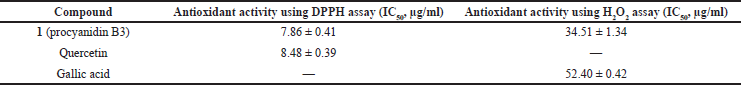
The antioxidant activity of the isolated compound was performed by the DPPH assay (Indrianingsih et al., 2021). The sample was diluted in methanol in several concentrations, reacted with DPPH (1.01 mM) at room temperature for 30 minutes in dark conditions. The absorbance of the final solution was measured using a UV spectrophotometer at 517 nm. The radical scavenging activity of compound 1 was calculated using

DPPH scavenging activity (%) = A 0 A 1 A 0 × 100 , (1)

where A0 is the absorbance of the control and A1 is the absorbance of the sample. The measurement was conducted in triplicate. Quercetin was used as the positive standard in the DPPH assay.

H2O2 assay

The capability of compound 1 in the H2O2 radical scavenging assay was evaluated according to the literature (Indrianingsih et al., 2015) with slight adjustments. H2O2 was diluted in phosphate buffer solution (PBS) (pH 7.4) to obtain the concentration of 40 mmol/l. Compound 1 (4 ml in Aquades) was reacted for 10 minutes with 0.6 ml of H2O2. The absorbance of the final solution was measured at 230 nm using a UV spectrophotometer.

Reducing power assay

According to the literature, the reducing power assay was conducted (Indrianingsih et al., 2015). Compound 1 (20 g/ml) in PBS (2.5 ml) was reacted with K3Fe(CN)6 (2.5 ml), left to stand for 20 minutes at 50°C, and added with trichloroacetic acid (2.5 ml). After centrifugation for approximately 8 minutes, the upper layer (2.5 ml) was reacted with Aquades and FeCl3 (0.5 ml). The final solution was evaluated at 700 nm using a spectrophotometer.

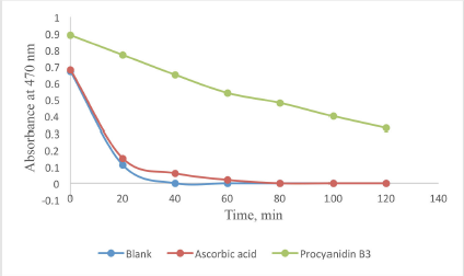
β-Carotene bleaching assay

According to a previous study, the capability of compound 1 in preventing β-carotene bleaching was performed (Indrianingsih et al., 2015). β-carotene (0.2 mg/ml) in CHCl3 was reacted with linoleic acid (20 mg) and Tween 40 (200 mg). After evaporation, distilled water (50 ml) was added, and 4.8 ml of the solution was reacted with 0.2 ml of compound 1 solution in methanol. This solution was incubated at 50C. The absorbance of the solution was evaluated at 20 minutes intervals at 470 nm.

α-Glucosidase inhibitory assay

The α-glucosidase assay was conducted according to the literature (Indrianingsih et al., 2015). Several concentrations of compound 1 in dimethyl sulfoxide (10 µl) were mixed with PBS (pH 7, 490 µl) and 250 µl of p-NPG (3 mmol/l). After being reacted for 5 minutes at 37°C, 250 µl of the α-glucosidase enzyme (0.065 IU/ml) was added and left to stand for 15 minutes. One milliliter of Na2CO3 (0.2 mol/l) was added to stop the reaction, and the absorbance of the final solution was evaluated at 400 nm.


RESULTS AND DISCUSSION

Isolation and structure identification

The chemical structure of compound 1 is illustrated in Figure 1. The ESI-MS of compound 1 recorded in positive ion mode exhibited a protonated ion [M + H]+ at m/z 579, indicating molecular formulas of C30H26O12. The 1H-NMR (Table 1) and 13C-NMR spectral data of compound 1 were compared with the literature (Kiss et al., 2008; Köhler et al., 2008; Wang et al., 2015), and structures were identified as procyanidin B3.

Figure 1. Compound 1 (procyanidin B3).

[Click here to view]

Several studies reported the bioactivity of procyanidins, such as antiproliferation (Kresty et al., 2011), antioxidant (Fu et al., 2013; Määttä-Riihinen et al., 2005), antimicrobial (Zang et al., 2013), and enzyme inhibitor (Wang et al., 2013) activities. In this study, the isolated procyanidin consisted of two catechin units; meanwhile, there are also existing procyanidins with five to six catechin or epicatechin units or larger polymeric procyanidins (Wang et al., 2015). However, the large polymeric procyanidins were little understood.

Antioxidant activity of compound 1

The antioxidant activities of compound 1 were evaluated using various assays: H2O2 radical scavenging activity, DPPH free radical scavenging activity, β-carotene-linoleate model assay, and reducing power assay. Table 2 shows the result of the DPPH assay and H2O2 assay.

Compound 1 showed the scavenging capacity against DPPH radicals (IC50) of 7.86 ± 0.38 µg/ml. Quercetin as control exhibited an IC50 of 8.48 ± 0.39 µg/ml. Based on this result, compound 1 has a higher antioxidant activity than quercetin. The good antioxidant activity of procyanidin B3 is probably due to the phenolic group of compound 1. A mechanism for the reaction was possible because the DPPH radical-driven catechin oxidation product attacks the A-ring unit, forms hydrophilic dimers, and oxidizes to oligomers (Osman, 2011). The DPPH assay was used for a simple, rapid, and efficient method (Kedar and Singh, 2011). Several studies showed that tocopherol, ascorbic acid, and phenolic compound could reduce DPPH radical form to its stable form (Arya et al., 2011; Liu et al., 2009).

Table 1. 1H-NMR (500 MHz, CD3OD) spectroscopic data for compound 1.

[Click here to view]

Table 2. The result of the DPPH and H2O2 assay of compound 1.

[Click here to view]

Figure 2. The result of the β-carotene bleaching assay of procyanidin B3 at 40 μg/ml.

[Click here to view]

Compound 1 also had scavenging abilities against hydroxide peroxide as shown in Table 2 (IC50) of 34.51 ± 1.34 µg/ml. The IC50 of gallic acid as control was 52.40 ± 0.42 µg/ml. The higher capability of compound 1 compared to gallic acid to scavenge free radicals showed that it had better potential as an antioxidant, in concurrence with the result from the DPPH assay. The ability evaluation of the isolated compound to scavenge H2O2 needs to be conducted since H2O2 could be toxic to the human body if it changed to the hydroxide radicals in cells (Kumar et al., 2012).

The β-carotene-linoleate bleaching assay was conducted to evaluate the isolated compounds’ ability to inhibit the bleaching caused by the peroxyl radical. The free radicals were formed when oxidized linoleic acid attacked the β-carotene molecules; thus, the β-carotene would undergo decolorization. This system consisted of an aqueous emulsion of linoleic acid and β-carotene that is similar to the food system that usually consists of water and lipid with some emulsifier. Figure 2 presents the ability of compound 1 (procyanidin B3) to prevent the decolorization of β-carotene.

The results showed that compound 1 efficiently prevented the oxidation of β-carotene in the system higher than that of ascorbic acid in the 120 minutes of the time range. Compound 1 retained antioxidant activity of 0.54% while ascorbic acid had 0.02% of activity after 60 minutes of assay; meanwhile, after 120 minutes, compound 1 still retained antioxidant activity of 0.33%, while ascorbic acid had 0% of activity. A study revealed that flavanols, anthocyanins, and phenolic acid were active in the β-carotene bleaching test (Singhal et al., 2014).

Figure 3 shows the result of the reducing power test of compound 1. This study used ascorbic acid and gallic acid equivalents to express the reducing power capability. It was presented that compound 1 had good reducing power activity of 87.80 mg/g [ascorbic acid equivalent (AAE)] and 50.49 mg/g [gallic acid equivalent (GAE)], in concurrence with H2O2 and DPPH radicals scavenging assays. The literature revealed that the antioxidant activity was related to the structure of a constituent, especially the active functional group (NH2 or OH). The position of the active group also had an important role, as the ortho position was more active than the para and meta position (Bendary et al., 2013).

α-Glucosidase inhibitor activity

Table 3 shows the IC50 values of α-glucosidase inhibitory activities of compound 1. Compound 1 had α-glucosidase inhibitory activity 58.81% ± 0.61% at a concentration of 50 µg/ml and IC50 of 40.05 ± 0.51 µg/ml. Meanwhile, the IC50 of quercetin was 4.20 ±0.01 µg/ml. A previous study reported that phenolic constituents had a strong inhibitory effect on α-glucosidase compared to acarbose (Zechel and Withers, 2000).

α-Glucosidase inhibitors play an essential role for DM patients with type 2 since they could inhibit the enzyme in the intestine and delay glucose absorption. Natural, safe, and effective α-glucosidase inhibitors from plants, microbes, and fungi were sought to develop functional food for DM patients (Katalinic et al., 2010). A study revealed that polyphenol could inhibit α-glucosidase and α-amylase since they also had good properties of antioxidant activities (Perron and Brumaghim, 2009). This study indicated that the isolated compound from Q. gilva leaves, procyanidin B3, has potential as the natural agent for type 2 DM patients since it is from nature. There is hope that pharmaceutical drugs for DM patients from nature have lower side effects than synthetic drugs (Eurich et al., 2007; Mohammed et al., 2013).

Figure 3. Antioxidant activities of procyanidin B3 using the reducing power assay. AAE: ascorbic acid equivalent; GAE: gallic acid equivalent.

[Click here to view]

Table 3. α-Glucosidase inhibitory activities of procyanidin B3 from Q. gilva.

[Click here to view]


CONCLUSION

In the present study, we have isolated one constituent (procyanidin B3) from the methanol extract of Q. gilva leaves. It showed satisfactory antioxidant activities using several assays such as the DPPH assay, H2O2 assay, β-carotene bleaching assay, and reducing power assay. The α-glucosidase inhibitory activity against the Saccharomyces cerevisiae α-glucosidase enzyme also showed promising results. This result will be helpful as important information for preparing pharmaceutical drugs sourced from nature.


AUTHOR CONTRIBUTIONS

All authors made substantial contributions to conception and design, acquisition of data, or analysis and interpretation of data; took part in drafting the article or revising it critically for important intellectual content; agreed to submit to the current journal; gave final approval of the version to be published; and agree to be accountable for all aspects of the work. All the authors are eligible to be an author as per the international committee of medical journal editors (ICMJE) requirements/guidelines.


FUNDING

There is no funding to report.


CONFLICTS OF INTEREST

The authors report no financial or any other conflicts of interest in this work.


ETHICAL APPROVALS

This study does not involve experiments on animals or human subjects.


DATA AVAILABILITY

All data generated and analyzed are included within this research article.


PUBLISHER’S NOTE

This journal remains neutral with regard to jurisdictional claims in published institutional affiliation.


REFERENCES

Arya V, Yadav S, Kumar S, Yadav JP. Antioxidant activity of organic and aqueous leaf extracts of Cassia occidentalis L. in relation to their phenolic content. Nat Prod Res, 2011; 25:1473–79. CrossRef

Bendary E, Francis RR, Ali HMG, Sarwat MI, Hady SE. Antioxidant and structure-activity relationships (SARs) of some phenolic and anilines compounds. Ann Agric Sci, 2013; 58(2):173–81. CrossRef

Brossa R, Casals I, Pintó-Marijuan M, Fleck I. Leaf flavonoid content in Quercus ilex L. resprouts and its seasonal variation. Trees, 2009; 23:401–8. CrossRef

Cantos E, Espín JC, López-Bote C, de la Hoz L, Ordóñez JA, Tomás-Barberán FA. Phenolic compounds and fatty acids from acorns (Quercus spp.), the main dietary constituent of free-ranged Iberian pigs. J Agric Food Chem, 2003; 51(21):6248–55. CrossRef

Deng J, Cheng W, Yang G. A novel antioxidant activity index (AAU) for natural products using the DPPH assay. Food Chem, 2011; 125(4):1430–5. CrossRef

Eurich DT, McAlister FA, Blackburn DF, Majumdar SR, Tsuyuki RT, Varney J, Johnson JA. Benefits and harms of antidiabetic agents in patients with diabetes and heart failure: a systematic review. BMJ, 2007; 335(7618):497. CrossRef

Fu CL, Wang HY, Ng WL, Song LX, Huang DJ. Antioxidant activity and proanthocyanidin profile of Selliguea feei rhizomes. Molecules, 2013; 18:4282–92. CrossRef

Gioti EM, Fiamegos YC, Skalkos DC, Stalikas CD. Antioxidant activity and bioactive components of the aerial parts of Hypericum perforatum L. from Epirus, Greece. Food Chem, 2009; 117:398–404. CrossRef

González-Palma I, Escalona-Buendía HB, Ponce-Alquicira E, Téllez-Téllez M, Gupta VK, Díaz-Godínez G, Soriano-Santos J. Evaluation of the antioxidant activity of aqueous and methanol extracts of Pleurotus ostreatus in different growth stages. Front Microbiol, 2016; 7:1099. CrossRef

Halliwell B. Free radicals and antioxidants: updating a personal view. Nutr Rev, 2012; 70(5):257–65. CrossRef

Indrianingsih AW, Tachibana S, Dewi RT, Itoh K. Antioxidant and α-glucosidase inhibitor activities of natural compounds isolated from Quercus gilva Blume leaves. Asian Pac J Trop Biomed, 2015a; 5(9):748–55. CrossRef

Indrianingsih AW, Wulanjati MP, Windarsih A, Bhattacharjya DK, Suzuki T, Katayama T. In vitro studies of antioxidant, antidiabetic, and antibacterial activities of Theobroma cacao, Anonna muricata and Clitoria ternatea. Biocatal Agric Biotechnol, 2021; 33:101995. CrossRef

Indrianingsih, AW, Tachibana S, Itoh K. In vitro evaluation of antioxidant and α-glucosidase inhibitory assay of several tropical and subtropical plants. Proc Environ Sci, 2015b; 28:639–48. CrossRef

Itokawa H, Tachi Y, Kamano Y, Iitaka Y. Structure of gilvanol, a new triterpene isolated from Quercus gilva Blume. Chem Pharm Bull, 1978; 26:331–3. CrossRef

Jakubczyk K, Ka?du?ska J, Dec K, Kawczuga D, Janda K. Antioxidant properties of small-molecule non-enzymatic compounds. Pol Merkur Lekarski, 2020; 48(284):128–32.

Jong J, Bimal K, Hyeun C, Kyung J, Ki S, Young S, Taek S, Ji L, Hye K, Min C. Comparison of phenolic compounds content in indeciduous Quercus species. J Med Plants Res, 2012; 6:5228–39. CrossRef

Katalinic V, Mozina SS, Skroza D, Generalic I, Abramovic H, Milos M, Ljubenkov I, Piskernik S, Pezo I, Terpinc P, Boban M. Polyphenolic profile, antioxidant properties and antimicrobial activity of grape skin extracts of 14 Vitis vinifera varieties grown in Dalmatia (Croatia). Food Chem, 2010; 119(2):715–23. CrossRef

Kedar SB, Singh RP. Genesis and development of DPPH method of antioxidant assay. J Food Sci Technol, 2011; 48(4):412–22. CrossRef

Kim H, You Y. Ecophysiological responses of Quercus gilva, endangered species and Q. glauca to long-term exposure to elevated CO2 concentration and temperature. J Ecol Environ, 2012; 35:203–12. CrossRef

Kiss AK, Derwi?ska M, Dawidowska A, Naruszewicz M. Novel biological properties of Oenothera paradoxa defatted seed extracts: effects on metallopeptidase activity. J Agric Food Chem, 2008; 56(17):7845–52. CrossRef

Klein O, Lynge J, Endahl L, Damholt B, Nosek L, Heise T. Albumin-bound basal insulin analogues (insulin detemir and NN344): comparable time-action profiles but less variability than insulin glargine in type 2 diabetes. Diabetes Obes Metab, 2007; 9(3):290–9. CrossRef

Köhler N, Wray V, Winterhalter P. Preparative isolation of procyanidins from grape seed extracts by high-speed counter-current chromatography. J Chromatogr A, 2008; 1177(1):114–25. CrossRef

Kozikowski AP, Tuckmantel W, Hu Y. Studies in polyphenol chemistry and bioactivity. 3.1,2 Stereocontrolled synthesis of epicatechin-4α,8-epicatechin, an unnatural isomer of the B-type procyanidins. J Org Chem, 2001; 66:1287–96. CrossRef

Kresty LA, Howell AB, Baird M. Cranberry proanthocyanidins mediate the growth arrest of lung cancer cells through modulation of gene expression and rapid induction of apoptosis. Molecules, 2011; 16:2375–90. CrossRef

Kumar RS, Rajkapoor B, Perumal P. Antioxidant activities of Indigofera cassioides Rottl. Ex. DC. using various in vitro assay models. Asian Pac J Trop Biomed, 2012; 2(4):256–61. CrossRef

Lämke JS, Unsicker SB. Phytochemical variation in treetops: causes and consequences for tree-insect herbivore interactions. Oecologia, 2018; 187(2):377–88. CrossRef

Liguori I, Russo G, Curcio F, Bulli G, Aran L, Della-Morte D, Gargiulo G, Testa G, Cacciatore F, Bonaduce D, Abete P. Oxidative stress, aging, and diseases. Clin Interv Aging, 2018; 13:757–72. CrossRef

Liu L, Sun Y, Laura T, Liang X, Ye H, Zeng X. Determination of polyphenolic content and antioxidant activity of kudingcha made from Ilex kudingcha CJ Tseng. Food Chem, 2009; 112:35–41. CrossRef

Lobo V, Patil A, Phatak A, Chandra N. Free radicals, antioxidants and functional foods: impact on human health. Pharmacogn Rev, 2010; 4(8):118–26. CrossRef

Määttä-Riihinen KR, Kähkönen MP, Törrönen AR, Heinonen IM. Catechins and procyanidins in berries of vaccinium species and their antioxidant activity. J Agric Food Chem, 2005; 53(22):8485–91. CrossRef

Mohammed SA, Yaqub AG, Sanda KA, Nicholas AO, Arastus W, Muhammad M, Abdullahi S. Review on diabetes, synthetic drugs and glycemic effects of medicinal plants. J Med Plants Res, 2013; 7(36):2628–37.

Moon MY, Baik JS, Kim SS, Jang WJ, Kim MS, Lee NH. Identification of antioxidative constituents from the branches of Quercus gilva Blume. J Soc Cosmet Sci Korea, 2009; 35:251–6.

Namkoong J, Kern D, Knaggs HE. Assessment of human skin gene expression by different blends of plant extracts with implications to periorbital skin aging. Int J Mol Sci, 2018; 19(11):3349. CrossRef

Noshiro S, Sasaki Y. Identification of Japanese species of evergreen Quercus and Lithocarpus (Fagaceae). IAWA J, 2011; 32:383–93. CrossRef

Oizumi Y, Mohri Y, Hirota M, Makabe H. Synthesis of procyanidin B3 and its anti-inflammatory activity. the effect of 4-alkoxy group of catechin electrophile in the Yb(OTf)(3)-catalyzed condensation with catechin nucleophile. J Org Chem, 2010; 75(14):4884–6. CrossRef

Olugbami JO, Gbadegesin MA, Odunola OA. In vitro free radical scavenging and antioxidant properties of ethanol extract of Terminalia glaucescens. Pharmacogn Res, 2015; 7(1):49–56. CrossRef

Osman AM. Multiple pathways of the reaction of 2,2-diphenyl-1-picrylhydrazyl radical (DPPH) with (+)-catechin: evidence for the formation of a covalent adduct between DPPH·and the oxidized form of the polyphenol. Biochem Biophys Res Commun, 2011; 412(3):473–8. CrossRef

Pandey A, Tripathi P, Pandey R, Srivatava R, Goswami S. Alternative therapies useful in the management of diabetes: a systematic review. J Pharm Bioallied Sci, 2011; 3(4):504–12. CrossRef

Perron NR, Brumaghim JL. A review of the antioxidant mechanisms of polyphenol compounds related to iron binding. Cell Biochem Biophys, 2009; 53(2):75–100. CrossRef

Petruk G, Del Giudice R, Rigano MM, Monti DM. Antioxidants from plants protect against skin photoaging. Oxid Med Cell Longev, 2018; 2018:1454936. CrossRef

Rodríguez-García CM, Ruiz-Ruiz JC, Peraza-Echeverría L, Peraza-Sánchez SR, Torres-Tapia LW, Pérez-Brito D, Tapia-Tussell R, Herrera-Chalé FG, Segura-Campos MR, Quijano-Ramayo A. Antioxidant, antihypertensive, anti-hyperglycemic, and antimicrobial activity of aqueous extracts from twelve native plants of the Yucatan coast. PLoS One, 2019; 14(3):e0213493. CrossRef

Singhal M, Paul A, Singh HP. Synthesis and reducing power assay of methyl semicarbazone derivatives. J Saudi Chem Soc, 2014; 18(2):121–7. CrossRef

Sitarek P, Merecz-Sadowska A, Kowalczyk T, Wieczfinska J, Zajdel R, ?liwi?ski T. Potential synergistic action of bioactive compounds from plant extracts against skin infecting microorganisms. Int J Mol Sci, 2020; 21(14):5105. CrossRef

Sukardiman, Ervina M. The recent use of Swietenia mahagoni (L.) Jacq. as antidiabetes type 2 phytomedicine: a systematic review. Heliyon, 2020; 6:e03536. CrossRef

Sy GY, Cissé A, Nongonierma RB, Sarr M, Mbodj NA, Faye B. Hypoglycaemic and antidiabetic activity of acetonic extract of Vernonia colorata leaves in normoglycaemic and alloxan-induced diabetic rats. J Ethnopharmacol, 2005; 98(1–2):171–5. CrossRef

Vinha A, Barreira J, Costa A, Oliveira M. A new age for Quercus spp. fruits: review on nutritional and phytochemical composition and related biological activities of acorns. Compr Rev Food Sci Food Saf, 2016; 15(6):947–81. CrossRef

Wang CM, Hsu YM, Jhan YL, Tsai SJ, Lin SX, Su CH, Chou CH. Structure elucidation of procyanidins isolated from Rhododendron formosanum and their antioxidative and anti-bacterial activities. Molecules, 2015; 20:12787–803. CrossRef

Wang HY, Song LX, Feng SB, Liu YC, Zuo G, Lai FL, He GY, Chen MJ, Huang DJ. Characterization of proanthocyanidins in stems of Polygonum multiflorum Thunb as strong starch hydrolase inhibitors. Molecules, 2013; 18:2255–65. CrossRef

Youn SH, Kwon JH, Yin J, Tam LT, Ahn HS, Myung SC, Lee MW. Anti-inflammatory and anti-urolithiasis effects of polyphenolic compounds from Quercus gilva Blume. Molecules, 2017; 22:1121. CrossRef

Zang X, Shang M, Xu F, Liang J, Wang X, Mikage M, Cai S. A-type proanthocyanidins from the stems of Ephedra sinica (Ephedraceae) and their antimicrobial activities. Molecules, 2013; 18(5):5172–89. CrossRef

Zechel DL, Withers SG. Glycosidase mechanisms: anatomy of a finely tuned catalyst. Acc Chem Res, 2000; 33:11–8. CrossRef

Reference

Arya V, Yadav S, Kumar S, Yadav JP. Antioxidant activity of organic and aqueous leaf extracts of Cassia occidentalis L. in relation to their phenolic content. Nat Prod Res, 2011; 25:1473-79. https://doi.org/10.1080/14786419.2010.545351

Bendary E, Francis RR, Ali HMG, Sarwat MI, Hady SE. Antioxidant and structure-activity relationships (SARs) of some phenolic and anilines compounds. Ann Agric Sci, 2013; 58(2):173-81. https://doi.org/10.1016/j.aoas.2013.07.002

Brossa R, Casals I, Pintó-Marijuan M, Fleck I. Leaf flavonoid content in Quercus ilex L. resprouts and its seasonal variation. Trees, 2009; 23:401-8. https://doi.org/10.1007/s00468-008-0289-5

Cantos E, Espín JC, López-Bote C, de la Hoz L, Ordóñez JA, Tomás-Barberán FA. Phenolic compounds and fatty acids from acorns (Quercus spp.), the main dietary constituent of free-ranged Iberian pigs. J Agric Food Chem, 2003; 51(21):6248-55. https://doi.org/10.1021/jf030216v

Deng J, Cheng W, Yang G. A novel antioxidant activity index (AAU) for natural products using the DPPH assay. Food Chem, 2011; 125(4):1430-5. https://doi.org/10.1016/j.foodchem.2010.10.031

Eurich DT, McAlister FA, Blackburn DF, Majumdar SR, Tsuyuki RT, Varney J, Johnson JA. Benefits and harms of antidiabetic agents in patients with diabetes and heart failure: a systematic review. BMJ, 2007; 335(7618):497. https://doi.org/10.1136/bmj.39314.620174.80

Fu CL, Wang HY, Ng WL, Song LX, Huang DJ. Antioxidant activity and proanthocyanidin profile of Selliguea feei rhizomes. Molecules, 2013; 18:4282-92. https://doi.org/10.3390/molecules18044282

Gioti EM, Fiamegos YC, Skalkos DC, Stalikas CD. Antioxidant activity and bioactive components of the aerial parts of Hypericum perforatum L. from Epirus, Greece. Food Chem, 2009; 117:398-404. https://doi.org/10.1016/j.foodchem.2009.04.016

González-Palma I, Escalona-Buendía HB, Ponce-Alquicira E, Téllez-Téllez M, Gupta VK, Díaz-Godínez G, Soriano-Santos J. Evaluation of the antioxidant activity of aqueous and methanol extracts of Pleurotus ostreatus in different growth stages. Front Microbiol, 2016; 7:1099. https://doi.org/10.3389/fmicb.2016.01099

Halliwell B. Free radicals and antioxidants: updating a personal view. Nutr Rev, 2012; 70(5):257-65. https://doi.org/10.1111/j.1753-4887.2012.00476.x

Indrianingsih AW, Tachibana S, Dewi RT, Itoh K. Antioxidant and α-glucosidase inhibitor activities of natural compounds isolated from Quercus gilva Blume leaves. Asian Pac J Trop Biomed, 2015a; 5(9):748-55. https://doi.org/10.1016/j.apjtb.2015.07.004

Indrianingsih AW, Wulanjati MP, Windarsih A, Bhattacharjya DK, Suzuki T, Katayama T. In vitro studies of antioxidant, antidiabetic, and antibacterial activities of Theobroma cacao, Anonna muricata and Clitoria ternatea. Biocatal Agric Biotechnol, 2021; 33:101995. https://doi.org/10.1016/j.bcab.2021.101995

Indrianingsih, AW, Tachibana S, Itoh K. In vitro evaluation of antioxidant and α-glucosidase inhibitory assay of several tropical and subtropical plants. Proc Environ Sci, 2015b; 28:639-48. https://doi.org/10.1016/j.proenv.2015.07.075

Itokawa H, Tachi Y, Kamano Y, Iitaka Y. Structure of gilvanol, a new triterpene isolated from Quercus gilva Blume. Chem Pharm Bull, 1978; 26:331-3. https://doi.org/10.1248/cpb.26.331

Jakubczyk K, Ka?du?ska J, Dec K, Kawczuga D, Janda K. Antioxidant properties of small-molecule non-enzymatic compounds. Pol Merkur Lekarski, 2020; 48(284):128-32.

Jong J, Bimal K, Hyeun C, Kyung J, Ki S, Young S, Taek S, Ji L, Hye K, Min C. Comparison of phenolic compounds content in indeciduous Quercus species. J Med Plants Res, 2012; 6:5228-39. https://doi.org/10.5897/JMPR12.135

Katalinic V, Mozina SS, Skroza D, Generalic I, Abramovic H, Milos M, Ljubenkov I, Piskernik S, Pezo I, Terpinc P, Boban M. Polyphenolic profile, antioxidant properties and antimicrobial activity of grape skin extracts of 14 Vitis vinifera varieties grown in Dalmatia (Croatia). Food Chem, 2010; 119(2):715-23. https://doi.org/10.1016/j.foodchem.2009.07.019

Kedar SB, Singh RP. Genesis and development of DPPH method of antioxidant assay. J Food Sci Technol, 2011; 48(4):412-22. https://doi.org/10.1007/s13197-011-0251-1

Kim H, You Y. Ecophysiological responses of Quercus gilva, endangered species and Q. glauca to long-term exposure to elevated CO2 concentration and temperature. J Ecol Environ, 2012; 35:203-12. https://doi.org/10.5141/JEFB.2012.025

Kiss AK, Derwi?ska M, Dawidowska A, Naruszewicz M. Novel biological properties of Oenothera paradoxa defatted seed extracts: effects on metallopeptidase activity. J Agric Food Chem, 2008; 56(17):7845-52. https://doi.org/10.1021/jf801372h

Klein O, Lynge J, Endahl L, Damholt B, Nosek L, Heise T. Albumin-bound basal insulin analogues (insulin detemir and NN344): comparable time-action profiles but less variability than insulin glargine in type 2 diabetes. Diabetes Obes Metab, 2007; 9(3):290-9. https://doi.org/10.1111/j.1463-1326.2006.00685.x

Köhler N, Wray V, Winterhalter P. Preparative isolation of procyanidins from grape seed extracts by high-speed counter-current chromatography. J Chromatogr A, 2008; 1177(1):114-25. https://doi.org/10.1016/j.chroma.2007.11.028

Kozikowski AP, Tuckmantel W, Hu Y. Studies in polyphenol chemistry and bioactivity. 3.1,2 Stereocontrolled synthesis of epicatechin4α,8-epicatechin, an unnatural isomer of the B-type procyanidins. J Org Chem, 2001; 66:1287-96. https://doi.org/10.1021/jo001462y

Kresty LA, Howell AB, Baird M. Cranberry proanthocyanidins mediate the growth arrest of lung cancer cells through modulation of gene expression and rapid induction of apoptosis. Molecules, 2011; 16:2375-90. https://doi.org/10.3390/molecules16032375

Kumar RS, Rajkapoor B, Perumal P. Antioxidant activities of Indigofera cassioides Rottl. Ex. DC. using various in vitro assay models. Asian Pac J Trop Biomed, 2012; 2(4):256-61. https://doi.org/10.1016/S2221-1691(12)60019-7

Lämke JS, Unsicker SB. Phytochemical variation in treetops: causes and consequences for tree-insect herbivore interactions. Oecologia, 2018; 187(2):377-88. https://doi.org/10.1007/s00442-018-4087-5

Liguori I, Russo G, Curcio F, Bulli G, Aran L, Della-Morte D, Gargiulo G, Testa G, Cacciatore F, Bonaduce D, Abete P. Oxidative stress, aging, and diseases. Clin Interv Aging, 2018; 13:757-72. https://doi.org/10.2147/CIA.S158513

Liu L, Sun Y, Laura T, Liang X, Ye H, Zeng X. Determination of polyphenolic content and antioxidant activity of kudingcha made from Ilex kudingcha CJ Tseng. Food Chem, 2009; 112:35-41. https://doi.org/10.1016/j.foodchem.2008.05.038

Lobo V, Patil A, Phatak A, Chandra N. Free radicals, antioxidants and functional foods: impact on human health. Pharmacogn Rev, 2010; 4(8):118-26. https://doi.org/10.4103/0973-7847.70902

Määttä-Riihinen KR, Kähkönen MP, Törrönen AR, Heinonen IM. Catechins and procyanidins in berries of vaccinium species and their antioxidant activity. J Agric Food Chem, 2005; 53(22):8485-91. https://doi.org/10.1021/jf050408l

Mohammed SA, Yaqub AG, Sanda KA, Nicholas AO, Arastus W, Muhammad M, Abdullahi S. Review on diabetes, synthetic drugs and glycemic effects of medicinal plants. J Med Plants Res, 2013; 7(36):2628- 37.

Moon MY, Baik JS, Kim SS, Jang WJ, Kim MS, Lee NH. Identification of antioxidative constituents from the branches of Quercus gilva Blume. J Soc Cosmet Sci Korea, 2009; 35:251-6.

Namkoong J, Kern D, Knaggs HE. Assessment of human skin gene expression by different blends of plant extracts with implications to periorbital skin aging. Int J Mol Sci, 2018; 19(11):3349. https://doi.org/10.3390/ijms19113349

Noshiro S, Sasaki Y. Identification of Japanese species of evergreen Quercus and Lithocarpus (Fagaceae). IAWA J, 2011; 32:383-93. https://doi.org/10.1163/22941932-90000066

Oizumi Y, Mohri Y, Hirota M, Makabe H. Synthesis of procyanidin B3 and its anti-inflammatory activity. the effect of 4-alkoxy group of catechin electrophile in the Yb(OTf)(3)-catalyzed condensation with catechin nucleophile. J Org Chem, 2010; 75(14):4884-6. https://doi.org/10.1021/jo1009382

Olugbami JO, Gbadegesin MA, Odunola OA. In vitro free radical scavenging and antioxidant properties of ethanol extract of Terminalia glaucescens. Pharmacogn Res, 2015; 7(1):49-56. https://doi.org/10.4103/0974-8490.147200

Osman AM. Multiple pathways of the reaction of 2,2-diphenyl1-picrylhydrazyl radical (DPPH) with (+)-catechin: evidence for the formation of a covalent adduct between DPPH•and the oxidized form of the polyphenol. Biochem Biophys Res Commun, 2011; 412(3):473-8. https://doi.org/10.1016/j.bbrc.2011.07.123

Pandey A, Tripathi P, Pandey R, Srivatava R, Goswami S. Alternative therapies useful in the management of diabetes: a systematic review. J Pharm Bioallied Sci, 2011; 3(4):504-12. https://doi.org/10.4103/0975-7406.90103

Perron NR, Brumaghim JL. A review of the antioxidant mechanisms of polyphenol compounds related to iron binding. Cell Biochem Biophys, 2009; 53(2):75-100. https://doi.org/10.1007/s12013-009-9043-x

Petruk G, Del Giudice R, Rigano MM, Monti DM. Antioxidants from plants protect against skin photoaging. Oxid Med Cell Longev, 2018; 2018:1454936. https://doi.org/10.1155/2018/1454936

Rodríguez-García CM, Ruiz-Ruiz JC, Peraza-Echeverría L, Peraza-Sánchez SR, Torres-Tapia LW, Pérez-Brito D, Tapia-Tussell R, Herrera-Chalé FG, Segura-Campos MR, Quijano-Ramayo A. Antioxidant, antihypertensive, anti-hyperglycemic, and antimicrobial activity of aqueous extracts from twelve native plants of the Yucatan coast. PLoS One, 2019; 14(3):e0213493. https://doi.org/10.1371/journal.pone.0213493

Singhal M, Paul A, Singh HP. Synthesis and reducing power assay of methyl semicarbazone derivatives. J Saudi Chem Soc, 2014; 18(2):121-7. https://doi.org/10.1016/j.jscs.2011.06.004

Sitarek P, Merecz-Sadowska A, Kowalczyk T, Wieczfinska J, Zajdel R, ?liwi?ski T. Potential synergistic action of bioactive compounds from plant extracts against skin infecting microorganisms. Int J Mol Sci, 2020; 21(14):5105. https://doi.org/10.3390/ijms21145105

Sukardiman, Ervina M. The recent use of Swietenia mahagoni (L.) Jacq. as antidiabetes type 2 phytomedicine: a systematic review. Heliyon, 2020; 6:e03536. https://doi.org/10.1016/j.heliyon.2020.e03536

Sy GY, Cissé A, Nongonierma RB, Sarr M, Mbodj NA, Faye B. Hypoglycaemic and antidiabetic activity of acetonic extract of Vernonia colorata leaves in normoglycaemic and alloxan-induced diabetic rats. J Ethnopharmacol, 2005; 98(1-2):171-5. https://doi.org/10.1016/j.jep.2005.01.024

Vinha A, Barreira J, Costa A, Oliveira M. A new age for Quercus spp. fruits: review on nutritional and phytochemical composition and related biological activities of acorns. Compr Rev Food Sci Food Saf, 2016; 15(6):947-81. https://doi.org/10.1111/1541-4337.12220

Wang CM, Hsu YM, Jhan YL, Tsai SJ, Lin SX, Su CH, Chou CH. Structure elucidation of procyanidins isolated from Rhododendron formosanum and their antioxidative and anti-bacterial activities. Molecules, 2015; 20:12787-803. https://doi.org/10.3390/molecules200712787

Wang HY, Song LX, Feng SB, Liu YC, Zuo G, Lai FL, He GY, Chen MJ, Huang DJ. Characterization of proanthocyanidins in stems of Polygonum multiflorum Thunb as strong starch hydrolase inhibitors. Molecules, 2013; 18:2255-65. https://doi.org/10.3390/molecules18022255

Youn SH, Kwon JH, Yin J, Tam LT, Ahn HS, Myung SC, Lee MW. Anti-inflammatory and anti-urolithiasis effects of polyphenolic compounds from Quercus gilva Blume. Molecules, 2017; 22:1121. https://doi.org/10.3390/molecules22071121

Zang X, Shang M, Xu F, Liang J, Wang X, Mikage M, Cai S. A-type proanthocyanidins from the stems of Ephedra sinica (Ephedraceae) and their antimicrobial activities. Molecules, 2013; 18(5):5172-89. https://doi.org/10.3390/molecules18055172

Zechel DL, Withers SG. Glycosidase mechanisms: anatomy of a finely tuned catalyst. Acc Chem Res, 2000; 33:11-8. https://doi.org/10.1021/ar970172

Article Metrics
44 Views 85 Downloads 129 Total

Year

Month

Related Search

By author names