INTRODUCTION
The electromagnetic spectrum covers the wavelengths (λ) of all existing electromagnetic radiation, from gamma radiation to radio waves. Within this broad spectrum, solar radiation includes ultraviolet radiation (UVR, 100–400 nm), visible light (VL, 400–760 nm), and infrared radiation (IR, 760–3,000 nm) (Sklar et al., 2013). It is important to mention that the solar radiation that reaches the surface of the earth depends on different factors, such as altitude, latitude, state of the ozone layer, environmental and atmospheric conditions, time of day, and seasons, which can increase or decrease its intensity and, therefore, the exposure of humans to it (Bais et al., 2015; McKenzie et al., 2003).
For the study of its biological effects, ultraviolet radiation is divided into three regions according to the wavelength range: Ultraviolet C (UVC) (100-290 nm), Ultraviolet B (UVB) (290-320 nm) and Ultraviolet A (UVA) (320-400 nm), the latter being subdivided into UVA II (320-340 nm) and UVA I (340-400 nm) (International Organization for Standardization, 2010). UVC is the most energetic UVR; however, its harmful effects in humans are considered negligible because it is absorbed almost entirely by the ozone layer; UVB constitutes 5%–10% of the UVR that reaches the earth and is less energetic than UVC but can cause direct cellular damage; finally, UVA corresponds to the remaining 90%–95% of the total UVR and has deeper penetration into the skin than UVB (Fig. 1) (Dalmau et al., 2018; Lorigo et al., 2018).
Repeated and prolonged exposure of the skin to UVA and UVB radiation can cause undesirable short-term and long-term biological effects, such as inflammation, erythema, direct and indirect DNA damage, oxidative stress, immunosuppression, pigmentation, and skin cancer (Sarkany, 2019). For this reason, the use of topical sunscreens, using chemical and physical sun filters as active ingredients, has been recommended and used as a preferred mechanism to counteract and/or protect against the harmful effects of UVR on the skin. However, the use of these ingredients is questioned in terms of efficacy and safety due to some factors, such as their degradation by photolysis, trigger contact, photocontact, and/or allergic dermatitis, presenting systemic absorption of themselves or their degradation products and thus generating systemic responses, such as acting as endocrine disruptors, allergen, or hapten, which has led to some withdrawals from the market (Jansen et al., 2013; Lorigo et al., 2018).
 | Figure 1. Percentage of penetration of the UV radiation into the skin and its biological effects. [Click here to view] |
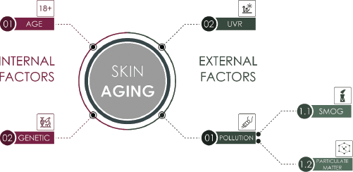
 | Figure 2. Factors that trigger skin aging. [Click here to view] |
On the other hand, one of the physiological states that is becoming increasingly important is skin aging, which is the result of the synergistic action of intrinsic and extrinsic effects on the structure and functionality of the skin (Fig. 2) (Farage et al., 2008). This has led in recent years to an increase in research of natural sources of photoprotective and antiaging bioactive compounds, to be used in the cosmetics industry to replace conventional sunscreens, as adjuvants to the same or other cosmetic products with the antiaging claim (Saewan and Jimtaisong, 2015).
This article reviews the interaction between UVR and molecules present in the skin, the biological effects that result from such interaction, the current state of sunscreens, and factors and mechanisms that trigger aging and also shows the potential cosmetic use of the bioactives present in marine macroalgae.
Interaction of UVR with biomolecules
UVR reaching the earth’s surface is capable of generating biological skin effects through its interaction with skin biomolecules. This interaction is based on the fact that UVR can be absorbed by chromophores, defined as molecules that are characterized by the presence of π-conjugated bonds. When a molecule absorbs UVR, it experiences changes in its electrons, because one of them is excited, going from a lower energy state to a higher energy state, but due to its instability in this new state, the molecule releases the energy either by the emission of fluorescence, phosphorescence, or heat, to return to the basal state (Gidolquim, 2016; Gilbertz et al., 2018). However, the recovery of the basal state in the molecule can also occur through chemical processes, where the molecule can undergo a chemical transformation involving radical dissociation, ion dissociation, and molecular reorganization. Besides, another mechanism called cross-systems may occur; in this case, the molecule transfers the absorbed energy to an unexcited molecule (Gidolquim, 2016).
In the skin, there are several important chromophores, such as DNA and intracellular and extracellular proteins, that undergo different functional and structural changes when absorbing UVR directly. However, these molecules can also be changed by the formation of reactive oxygen species (ROS), such as the hydroxyl radical (OH•), hydroperoxide radical (HO2•), superoxide radical (•O2−), and singlet oxygen (1O2) (Pattison et al., 2012). ROS can be formed either in the process of direct absorption of UVR by biomolecules or through type I and type II photoreactions, involving endogenous molecules such as cytochromes, flavins, heme, and porphyrins, among others, known as sensitizers, by absorbing UVR transfer excitation energy to an intermediate molecule through the transfer of electrons or hydrogen atoms (type I) or directly to molecular oxygen in a triplet state (type II). In this sense, ROS are considered second messengers of UVR, due to the fact that they can cause harmful biological effects through their action on skin biomolecules (Cadet et al., 2015; Da et al., 2017).
Action spectrum
It is important to mention that the biological effects triggered by UVR on the skin are wavelength-dependent; i.e., not all wavelengths are harmful or beneficial to the same extent since there are some of them where the biological effect obtained is maximum (Bais et al., 2015). The biological action spectra are graphs wavelength versus reciprocal of radiation that allows knowing the effectiveness of different wavelengths of radiation to generate a biological effect, thus allowing to determine the incident wavelength that is capable of producing a specific response, defined as effective biological UV irradiance (UVeff) and obtained by multiplying the spectrum of solar radiation with the action spectrum for the biological effect under study. Due to the effects of UVR on the skin, the spectra of erythematous action, DNA absorption of UVR, and non-melanoma skin cancer are considered the most important and used in the evaluation of the biological effects produced by UVR (Bais et al., 2015; De Argila et al., 2014; De Fabo, 2006; De Gruijl, 2000; Matts, 2006). One of the concepts derived from the erythematous action spectrum is the minimum erythema dose minimal erythema dose (MED), which refers to the minimum amount of UVR capable of producing the first noticeable and unambiguous erythema (redness of the skin), with defined edges, without previous exposure, and is determined 16–24 hours after exposure to UVR; it can also be defined as the amount of energy required per unit area (J/cm2) for the appearance of minimum erythema (International Organization for Standardization, 2010).
Skin structure
The skin is the most extensive organ of the body. It is composed of three layers: epidermis, dermis, and hypodermis. The epidermis is the most external layer; it is metabolically active because it periodically has a renewal process; its main cells are the keratinocytes, which after being submitted to different biochemical and morphological changes give rise to the formation of the stratum corneum which is considered the fundamental physical barrier of the skin. There are also the melanocytes in charge of producing melanin, the Langerhans cells that participate in the immune function of the skin, and the Merkel cells that act as sensory receptors (James et al., 2020). The dermis is the underlying layer of the epidermis; the fibroblasts are the main cells, but they also contain a large extracellular matrix (ECM) which is composed of molecular structures such as structural proteins, including collagen and elastin, as well as macromolecules that participate in different biological processes of the skin such as hyaluronic acid, a polysaccharide that provides hydration (D’Orazio et al., 2013; Gu et al., 2020; James et al., 2020). The molecular structures present in the epidermis and dermis of the skin are those that interact with the UVR, thus generating the different biological effects. It is important to mention that UVA radiation can penetrate the dermis, while UVB radiation is mostly absorbed in the epidermis (Fig. 1) (Samaniego Rascón et al., 2017).
Biological effects of UVR
Low-dose UVR induces the conversion of 7-dehydrocholesterol to vitamin D, which is important in calcium binding in the bone system (Fioletov et al., 2009; Sarkany, 2019). However, excessive exposure to UVR is a health risk, due to the generation of acute and chronic effects (D’Orazio et al., 2013). Acute effects include sunburn (erythema), which can be mild or severe; epidermal thickening (hyperkeratosis), mainly of the stratum corneum; immunosuppression and skin pigmentation (tanning) that involve immediate darkening of the pigment immediate pigment darkening (IPD), persistent darkening of the pigment, permanent pigment darkening (PPD), and late tanning or delayed tanning (DT), which appears according to the amount of radiation received; furthermore, they differ in that the IPD usually appears seconds after exposure, the PPD appears 2–24 hours later, and the DT can be observed 72 hours after exposure. On the other hand, the chronic effects of UVR are photoaging (dry, wrinkled, and inelastic skin) and skin cancer, which is divided according to the type of cell affected into melanoma and nonmelanoma, the latter being subdivided into both basal cell and squamous cell carcinoma (Matsumura and Ananthaswamy, 2004; Poon et al., 2014; Samaniego Rascón et al., 2017; Sarkany, 2019). UVR can also cause eye diseases such as cataract, photokeratitis, and tumor formation, among others, through interaction with molecules present in the structure of the eye (Yam and Kwok, 2014). Figure 1 shows the percentage of penetration of UVR into the skin and its biological effects.
To protect against the effects of UVR, the skin contains several photoprotective molecules capable of absorbing radiation, including urocanic acid, melanin, bilirubin, reduced hemoglobin, and aromatic amino acids, such as tryptophan and tyrosine (Jansen et al., 2013; Juzeniene et al., 2009; Young et al., 2017). Also, the skin has an enzymatic and nonenzymatic defense system against ROS caused by UVR. The first group includes the enzymes catalase, glutathione peroxidase, and superoxide dismutase, while the second group includes antioxidants such as glutathione (Kullavanijaya and Lim, 2005). However, with prolonged exposure to UVR, these mechanisms can be exhausted and because of this, the use of sunscreens is necessary to counteract the effects of UVR on the skin.
Photoprotection: sun filters
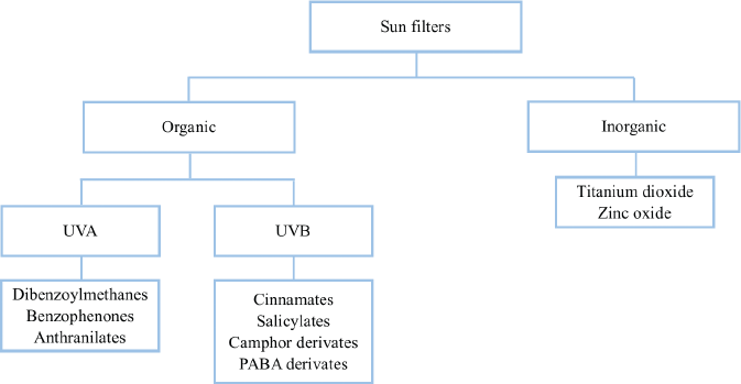
A sunscreen or UV filter is defined by the Food and Drug Administration (FDA) as an active ingredient that absorbs, reflects, or scatters UVR at wavelengths of 290–400 nm (Food and Drug Administration, 2011). However, a sunscreen must also be stable, safe, preferably broad spectrum, and possess acceptable sensory characteristics (Geoffrey et al., 2019; Jansen et al., 2013). The UV filters available in the market and therefore in commercial sunscreens are classified into two groups: organic or chemical filters and inorganic or physical filters, which differ basically in their chemical structure and mechanism of action; the former are aromatic compounds that absorb UVR and the latter are minerals that reflect, disperse, and/or absorb UVR (Mancebo et al., 2014). Figure 3 shows the classification of sun filters.
Furthermore, it is possible to classify them according to the UV range in which they absorb as UVB filters, UVA filters, or broad-spectrum filters, which absorb both UVB and UVA radiation (Osterwalder et al., 2014).
Organic filters are molecules that are characterized by having in their structure one or more aromatic rings (conjugated electrons π), with substitutions in position para or ortho of electron donor groups, besides, they have hydrophobic groups that improve their properties, such as their substantivity to the skin (Cadena-Aizaga et al., 2020; Jansen et al., 2013; Osterwalder et al., 2014). According to their UVR absorption range and the chemical group they are divided into UVA filters (dibenzoylmethanes, benzophenones and anthranilates) and UVB filters (cinnamates, salicylates, camphor derivatives and Para Aminobezoic Acid (PABA) derivatives) (Cadena-Aizaga et al., 2020; Osterwalder et al., 2014; Wong and Orton, 2011). Organic UV filters exert their photoprotective action by absorbing UVR, which causes that one electron in the molecule jump into an excited state and when it returns to its fundamental state, it releases the absorbed energy through different mechanisms such as heat or at longer wavelengths (fluorescence; phosphorescence) (Mancebo et al., 2014). It is important to mention that each organic UV filter has an absorption spectrum limited to a range of wavelengths, which is why sunscreen formulations contain an association (Cadena-Aizaga et al., 2020; Kockler et al., 2012).
 | Figure 3. Classification of sun filters. It is adapted from Geoffrey et al. (2019). [Click here to view] |
Inorganic UV filters are chemically inert minerals that act as a barrier by absorbing, scattering and reflecting UVR in the range of 290-400nm, and therefore they are considered as broad-spectrum filters. Among this group, the two filters approved for use in sunscreens are titanium dioxide (TiO2) and zinc oxide (ZnO) (Kockler et al., 2012). The reflection and scattering capacity of these filters depend largely on particle size, so large particle sizes (larger than nanometers scale) with high light reflection index leave a white film on the skin surface. For this reason, the cosmetic industry has opted for reducing particle size to nanometers or micrometers, in order to decrease light reflection and improve not only consumer acceptability but also UVR absorption, reflection, and scattering. These filters in comparison with the chemical filters have a lesser instability by the UVR; however, being semiconductor materials, they can generate ROS; nevertheless, this effect has been improved using its nanoencapsulation with aluminum oxide or silica mainly, which avoids the adhesion of the filter to the skin and, therefore, the passage of ROS to the same one (Kockler et al., 2012; Mancebo et al., 2014; Osterwalder et al., 2014).
Nevertheless, in the mechanism of action of organic UV filters, there are some photoreactions derived from their interaction with UVR, which lead to structural changes in the molecule, for example, trans-cis isomerization in ethylhexyl methoxycinnamate (Hojerová et al., 2011; Mancebo et al., 2014), and several studies have shown that butyl methoxydibenzoylmethane is one of the most photounstable filters due to its keto-enol tautomerism and fragmentation with the formation of photolysis products (Chatelain and Gabard, 2001; Gaspar and Maia Campos, 2006; Karlsson and Hillerström, 2009). These phenomena cause the loss of effectiveness and safety of these molecules. For these reasons, the photostability of sunscreens is an important parameter to evaluate in their development, since it allows establishing the stability of the filters by interacting with the UVR, a factor that in turn determines their effectiveness and safety (Berkey et al., 2019). Nowadays, sunscreens are the most common cause of contact, photocontact, phototoxic, and photoallergic dermatitis around the world (Bryden et al., 2006; Kockler et al., 2012; Victor et al., 2010; Wong and Orton, 2011). In the 1990s, benzophenone-3 was the most common photoallergenic sunscreen and isopropyldibenzoylmethane was withdrawn from the market in 1993, after being identified as the most common cause of photoallergic contact dermatitis in the 1980s–1990s (Wong and Orton, 2011). Therefore, it is necessary to improve the effectiveness of sunscreens through new sources of photoprotective compounds that can replace or reduce the concentrations of traditionally used filters. Thus, various studies have been carried out on groups of secondary metabolites and extracts from seaweed, in order to evaluate their possible photoprotective properties and obtain new sources of raw materials of natural origin.
Efficacy test on sunscreens
Sunscreens are considered as medicine in the United States of America and the United Kingdom, while in the European Union, they are part of cosmetics. Based on this, the efficacy tests for these products are carried out using in vitro and in vivo methodologies according to the protocols of each regulatory entity. For example, in the USA, the protocols established by the FDA are applied, but in the United Kingdom, the Boot’s star system is used and in the countries of Europe, the protocols given by COLIPA (The European Cosmetic, Toiletry and Perfumery Association) and International Organization for Standardization (ISO) are used (Geoffrey et al., 2019; Kaimal and Abraham, 2011; Osterwalder et al., 2014).
Tests in vivo
One of the in vivo methods to evaluate the efficacy of sunscreens is the determination of the sun protection factor (SPF). SPF is considered the universal indicator of the effectiveness of a sunscreen to protect from UVB radiation because it is calculated by dividing the MED dose of the skin protected with the test product by the MED dose of the unprotected skin (European Commission, 2006). This method that is carried out by exposing human subjects to a source of artificial UVR does not therefore indicate that the evaluated sunscreen protects against UVA radiation, since erythema (redness) or sunburns are a biological effect caused mainly by wavelengths of 290–320 nm, with maximum absorption at 308 nm, which corresponds to the UVB range (European Commission, 2006; FDA, 2011; International Organization for Standardization, 2010).
Therefore, the PPD method is a method that allows evaluating the ability of a sunscreen to protect against UVA radiation. Like SPF, it is carried out on human subjects exposed to UVR from an artificial source and allows calculating the UVA protection factor (UVA-PF) by dividing the minimum pigmentation dose or minimal pigmenting dose (MPD) on the skin protected by MPD on unprotected skin. MPD is the smallest dose of UVA energy that causes minimal unambiguous pigmentation with bounded edges after 2–4 hours of radiation exposure as a result of melanin photooxidation reactions. However, it is necessary to mention that, due to human exposure to UVA radiation in carrying out this method, in addition to factors such as cost and time, there are alternative in vitro methods that allow obtaining results equivalent to those obtained with in vivo methodologies alive (European Commission, 2006; FDA, 2011).
Tests in vitro
For the evaluation of the in vitro protective capacity of a sunscreen against UVA radiation, there are three proposed methodologies: one from the USA through the FDA, one from Europe available in ISO 24443 in 2012, and another from the United Kingdom known as the Boot's star system, which preferably uses the transmittance technique.
The FDA proposes the critical wavelength (λc) method, defined as the wavelength where the area under the curve of the absorbance spectrum for the irradiated product (obtained by FDA methodology) from 290 nm corresponds to 90% of the total area of the absorbance spectrum of 290–400 nm. The FDA establishes a critical wavelength greater than or equal to 370 nm as a criterion for the declaration of a broad spectrum in the sunscreens under study (FDA, 2011, 2012).
Meanwhile, the European Union not only considers the critical wave methodology to measure the UVA protection capacity of the product under study but also proposes the determination of the UVA protection factor in vitro, since it has been shown to correlate with the obtained in vivo tests in the PPD method. Therefore, the European Union establishes two parameters for the broad spectrum declaration: λc ≥ 370 nm and UVA-PF≥1/3 of the SPF value obtained for the product in in vivo test (European Commission, 2006; International Organization for Standardization, 2012). Lastly, the method proposed in the United Kingdom known as the Boot’s star system is based on the calculation of the relationship between the absorbances obtained in the UVA and UVB regions, measured before and after the application of the photoprotector under study. This system allows a star rating, ranging from zero to five, where the higher the ratio, the greater the number of stars and the greater the protection of the product against UVA radiation (Kaimal and Abraham, 2011; Padera, 2011).
Regarding the determination of the SPF in vitro, several methodologies have been developed, which are sometimes used and supported by in silico measurements in the process of developing sunscreens; however, there is no standardized or harmonized methodology by regulatory entities for the in vitro determination of this parameter (Osterwalder and Herzog, 2009; Stanfield et al., 2010). Table 1 describes the main characteristics of protocols for evaluating the effectiveness of sunscreens.
Skin aging
Skin aging is a degenerative biological process that results from the action of intrinsic and extrinsic factors on skin tissue. Figure 2 shows the factors that trigger skin aging. Among the intrinsic factors are mainly age and genetics, while extrinsic factors include environmental pollution (smog, particulate matter, smoke, etc.) and UVR, which is considered as the extrinsic factor that mostly induces aging. These factors exert a synergistic effect on each other that produces a loss of the integrity and functionality of the skin that is reflected through a series of clinical manifestations. However, there are differences between these, when aging corresponds to the natural physiological process and when it is due to external factors (Farage et al., 2008; Fussell and Kelly, 2020).
Intrinsic aging is characterized by a thinning of the epidermis due to a decrease in the ability of keratinocytes to proliferate and renew, in addition to reduction not only of epidermal stem cells but also of the lipid synthesis of the stratum corneum; as for the thickness of the dermis, it also decreases because the number of fibroblasts decreases and with it the synthesis of collagen and elastin decreases; these alterations lead to dry, smooth skin with fine wrinkles, less elastic and sometimes benign neoplasms (Strnadova et al., 2019). On the other hand, extrinsic aging is characterized by an increase in the thickness of the epidermis; in the dermis, the number of fibroblasts is less, the synthesis of the components of the ECM decreases, and there is an accumulation of disorganized amorphous collagen and elastin fibers as a consequence of an increase in their degradation. Unlike intrinsic aging, extrinsic aging results in roughly textured, unevenly pigmented skin, with a yellowish color, thick and deep wrinkles, and very little elasticity, with telangiectasias (dilation of small blood vessels) and benign and malignant neoplasms can occur (Gruber et al., 2020; Landau, 2007; Strnadova et al., 2019).
 | Table 1. Summary of the main characteristics of protocols for evaluating the effectiveness of sunscreens. [Click here to view] |
Despite the differences between the clinical manifestations of intrinsic and extrinsic aging, both are the result of alterations at the cellular and molecular level caused by the aforementioned factors. Among these alterations, one of the most important alterations involves the components of the dermal ECM, especially the organized fibers of collagen and elastin, which play an important role in the mechanical resistance and elasticity of the skin. In the aging process of the skin, there is a decrease in these proteins through two mechanisms, the first is by the inhibition of the synthesis pathways and the second by stimulating the expression of matrix metalloproteinases (MMPs), a family of zinc-containing proteolytic enzymes whose function is to degrade the molecular structures present in the ECM; their overproduction leads to an increase in disorganized fragments of collagen and elastin in the connective tissue, resulting in elastosis, characterized by the skin with wrinkles and thick, as well as other clinical manifestations of aging (Quan et al., 2004, 2009). The main MMPs involved in this mechanism are MMP-1 (collagenase) which acts on type I collagens (majority collagen in ECM) of types II, III, VII, and X; MMP-3 (stromelicin-1) whose substrates are elastin and collagens of types IV, V, IX, and X. Furthermore, MMP-9 (gelatinase) is also involved, which degrades mainly elastin and types I, IV, and V collagens (Pérez-García, 2004; Quan et al., 2004).
On the other hand, there are also ROS that have an important role in skin aging, both intrinsic and extrinsic. ROS can be the result of normal endogenous biological processes and the action of exogenous factors; in the first case, they are generated in the electron transport chain in the aerobic metabolic process of the skin; an example of this is the formation of the superoxide radical in the oxidation of NADPH to NADP+ by the enzyme NADPH oxidase; however, this and other ROS are also mainly generated by the action of UVR through the aforementioned mechanisms (Fisher et al., 2009; Gu et al., 2020). ROS participates in aging through different mechanisms: they can alter gene expression and signaling pathways, activate matrix metalloproteinase transcription, and expression by activating the AP-1 nuclear transcription complex and inactivating MMP tissue inhibitors. They cause cellular lipids oxidation, damage to proteins through the oxidation of amino acids, and damage to cellular and mitochondrial DNA, through the oxidation of guanine to 8-oxoguanin, which can, in turn, cause damage to the telomeres that participate in cell proliferation and whose decrease is considered a marker of aging (Fussell and Kelly, 2020; Gu et al., 2020; Kosmadaki and Gilchrest, 2004; Tobin, 2017).
All these mechanisms contribute to skin aging either intrinsically or extrinsically; however, hydrated, nourished, and protected skin against the effects of UV radiation has a greater resistance to them.
Potential photoprotective and antiaging molecules
Like humans, marine macroalgae are affected by high exposure to UVR, as well as other extreme environmental conditions of pH, temperature, and high salinity that have led to the development of defense mechanisms against these factors, such as the synthesis of bioactive compounds. Among these compounds are mycosporine-type amino acids (MAAs), polyphenols, and sulfated polysaccharides, among others, which have been shown to have various biological activities, such as photoprotective, antioxidant, and anti-inflammatory capacity, among others, on the skin (Pangestuti et al., 2018; Wang et al., 2015).
Mycosporine-type amino acids
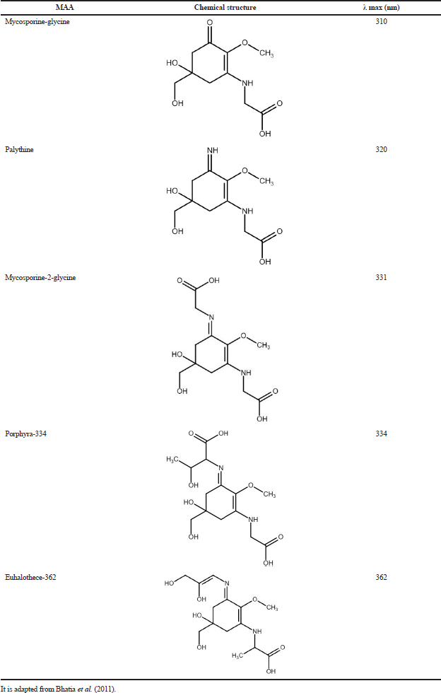
Mycosporine type amino acids (MMAs) are secondary metabolites synthesized as a defense mechanism against the effects of solar radiation. It is characterized by having a molecular weight less than 400 Da and a ring of cyclohexenone or cycloheximide (chromophore group) with nitrogenous substituents, mainly amino acid residues and amino alcohols (Bhatia et al., 2011; Kageyama and Waditee-Sirisattha, 2019). The MAAs can be divided according to the number of substitutions in monosubstituted and disubstituted: the former has substitutions in carbon three (C3) and among them is mycosporine-glycine which is considered as an intermediate MAAs from which disubstituted MAAs are found (mainly substituted in C1 and C3). MAAs have absorption in the UVB and UVA radiation range (310–360 nm) with molar absorption coefficients between 28,000 and 50,000 M−1cm−1; however, their absorption maxima depend on the ring substituents; for example, mycosporine-glycine has a maximum absorbance at 310 nm, while porphyria 334 substituted with threonine and glycine has an absorption maximum at 334 nm. An important feature in MMAs is its photostability, which will then absorb radiation and release energy in the form of heat upon returning to the ground state (Kageyama and Waditee-Sirisattha, 2019; Lalegerie et al., 2019). Table 2 shows different mycosporine amino acids with the chemical structures and absorption maxima.
Sulfated polysaccharides
The main sulfated polysaccharides in red macroalgae are carrageenans; meanwhile, fucoidans are present in brown algae (Pangestuti et al., 2018). Carrageenans are composed of galactose and anhydrogalactose units that repeat with alternations of α (1→4) and ß (1→3) type, that is, 3-linked β-D-galactopyranose and 4-linked α-galactopyranose or 3–6-anhydro-α-galactopyranose. In these molecules, the sulfate group in the galactose unit can be in C4 and C2, while in the 3–6-anhydro-α-galactose unit, it can be in C2, C1, and C6, or it may not be sulfated (Jiao et al., 2011). Fucoidan, on the other hand, is characterized by being polysaccharides with sulfated L-fucose in a greater proportion than other saccharides that can also be found as galactose, urocanic acids, mannose, and xylose. In fucoidans, the sulfated portion can be found in C2, C3, or C4 and monosaccharides are associated through α (1→2), α (1→3), or α (1→4) glycosidic bonds; their size can vary from 13 to 950 kDa (Berteau and Mulloy, 2003; Holtkamp et al., 2009).
 | Table 2. Chemical structures of different mycosporine amino acids with absorption maxima (nm). [Click here to view] |
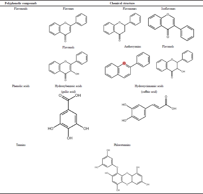
 | Table 3. Classification of polyphenolic compounds with chemical structures. [Click here to view] |
Polyphenols
Polyphenolic compounds are a broad group of secondary metabolites that are characterized by having one or more mono- or polysubstituted aromatic rings in their structure by the hydroxyl group which is considered as an electron donor (Pangestuti et al., 2018). Polyphenols include flavonoids, phenolic acids, and tannins. Table 3 shows the classification of polyphenolic compounds with chemical structure.
Flavonoids are characterized by having a C6–C3–C6 configuration for a total of 15 carbons; they have two aromatic rings called A and B, which are united through a chain of three carbons, which is generally found to form a heterocyclic ring (ring C), which according to the substitutions it presents allows obtaining six subgroups: anthocyanins, flavones, isoflavones, flavonols, flavanols, and flavanones. Phenolic acids are characterized by having a carboxyl group; they are subdivided into hydroxycinnamic acids with a C6–C3 configuration and hydroxybenzoic acids with a C6–C1 configuration (Ignat et al., 2011). Tannins can be divided into hydrolyzable, condensable, and phlorotannins; the latter are mainly associated with brown algae (Balboa et al., 2013; Ignat et al., 2011).
The presence of these compounds in marine macroalgae has drawn attention in research intending to determine the biological activities derived from them. For example, Souza et al. (2011) studied two species of red macroalgae on the coasts of Brazil and found that the ethanolic and methanolic extracts showed antioxidant effects due to the presence of polyphenolic compounds, such as gallic acid and apigenin (Souza et al., 2011). On the other hand, Kim et al., 2018, evaluated the anti-photoaging capacity of a fucoidan and concluded that this sulfated polysaccharide had an antioxidant effect and inhibited the expression of MMPs and confirmed several studies on macroalgae extracts (Kim et al., 2018, 2006; Ryu et al., 2009). Also, its photoprotective effect has been evaluated; for example, Bhatia et al. (2019) developed a formulation with MAAs from the macroalgae Ulva fasciata with an SPF 7, a value that correlates with that found by Álvarez-Gómez et al. (2019), when evaluating the in vitro photoprotective capacity (SPF 7.5) of the red macroalgae Gracilariopsis longissima.
Based on these results, studies have been carried out on the use of macroalgae extracts in photoprotective and antiaging cosmetic formulations, for which the process of collection and extraction of bioactive compounds, as well as their isolation and purification, has been necessary.
Seaweeds are collected in coastal areas and are subjected to a washing process to eliminate impurities. Then, they are dried in an oven or the air and made to a fine powder by crushing to continue the extraction process (Bhatia et al., 2019; Bittkau et al., 2020; Poulose et al., 2020). Currently, there are several techniques available for the extraction of bioactive compounds present in macroalgae, among which are conventional solid–liquid extraction (SLE), ultrasound-assisted extraction, microwave-assisted extraction, enzymes assisted extraction, and supercritical fluid extraction; however, due its simplicity and easy scalability, SLE is the most widely used (Pangestuti et al., 2018; Santos et al., 2019).
It is important to mention that the extraction conditions may vary according to the bioactive compounds of interest; for example, for the extraction of MAAs, aqueous binary mixtures with ethanol and methanol are used as solvents (Bhatia et al., 2019; Ryu et al., 2014; Zhaohui et al., 2005); although the proportions may be different, some authors have agreed that the temperature and time of extraction are 45ºC for 2 hours (Hartmann et al., 2016; Hoyer et al., 2002). On the other hand, for the extraction of phenolic compounds as in the extraction of MAAs aqueous binary mixtures with ethanol, methanol, acetone, and acetonitrile at different concentrations, times, and temperatures are used (Santos et al., 2019). Vu et al. (2017) evaluated the content of phenolic compounds in the macroalgae Sargassum serratum at various conditions and concluded that the highest content of polyphenols and antioxidant activity was obtained when extraction was performed with 100% ethanol at 50ºC for 32 hours. In the case of sulfated polysaccharides, the extraction can be carried out using hot water or acidified solutions as solvents at room temperature; however, a key process in the extraction of sulfated polysaccharides is the addition of CaCl2 to avoid the coextraction of alginic acid (Bittkau et al., 2020; Kim et al., 2007). Notwithstanding, the yield obtained in the extraction of macroalgae depends not only on the bioactive compounds of interest, technique used, and extraction conditions, but also on the species of algae evaluated.
After the extraction of bioactive compounds from macroalgae, their purification can be obtained by different techniques; for example, Hartman et al. (2016), obtained an extract enriched in MAAs from Prasiola calophylla through an adsorption-based separation technique using an ion exchange resin and monitored by preparative high-performance liquid chromatography (HPLC); the purified extract was analyzed by nuclear magnetic resonance, which allowed the identification of MAA N-[5,6 hydroxy5(hydroxymethyl)-2-methoxy-3-oxo-1-cycohexen-1-yl] glutamic acid (prasiolin) as a new photoprotective compound present in macroalgae (Hartmann et al., 2016). Nevertheless, Bhatia et al., 2019 purified the ethanolic extract from Ulva fasciata using a silica gel column and monitoring by Thin Layer Chromatography (TLC), subsequently, the purified fraction was analyzed by HPLC with Photodiode Array (PDA) detector and Liquid Chromatography-Mass Spectrometry (LC-MS). Another technique used for the purification of extracts obtained from macroalgae is molecular-weight cut-off dialysis (MWCO). Kim et al. (2007) purified the extract obtained from Undaria pinnatifida by means dialysis (MWCO 14,000) and column chromatography using a cellulose column, from which they obtained a fraction rich in sulfated polysaccharides; then, they analyzed using high performance anion-exchange chromatography with pulsed amperometric detector.
Additionally, studies have been carried out on the use of macroalgae extracts in photoprotective and antiaging cosmetic formulations. For example, Mercurio et al. (2015) mixed UV filters together with Porphyra umbilicalis (red algae) extract and found that this combination not only improved the photoprotective capacity of the formulation but also had a positive effect on cell proliferation; results indicated that the bioactive compounds in the macroalgae have potential use for the development of photoprotective and antiaging cosmetic formulations (Mercurio et al., 2015).
CONCLUSION
In recent years, research studies have grown into new natural sources of molecules with potential photoprotective and antiaging activity that is safe and effective. Among these sources, one of the most promising sources is marine macroalgae, due to the environmental conditions in which they are found; they have generated mechanisms of protection against radiation, such as the synthesis of secondary metabolites with photoprotective, antioxidant, anti-inflammatory, and antiaging activity. For this reason, it makes them potential active compounds in the cosmetic industry for the development of products with a photoprotective and antiaging effect, the latter being useful to counteract the action of intrinsic and extrinsic factors on skin aging.
ACKNOWLEDGMENTS
Luna Pérez Y. acknowledges master fellowship granted by University of Antioquia (Programa de Becas a Mejor Graduado). This review is part of the project granted by CODI-University of Antioquia (Project no. 2019-25210).
CONFLICT OF INTEREST
The authors declare that they have no conflicts of interest among them or with the parent institution.
AUTHOR CONTRIBUTIONS
All authors made substantial contributions to conception and design, acquisition of data, or analysis and interpretation of data; took part in drafting the article or revising it critically for important intellectual content; agreed to submit to the current journal; gave final approval of the version to be published; and agree to be accountable for all aspects of the work.
REFERENCES
Álvarez-Gómez F, Korbee N, Casas-Arrojo V, Abdala-Díaz RT, Figueroa FL. UV photoprotection, cytotoxicity and immunology capacity of red algae extracts. Molecules, 2019; 24:1–16. CrossRef
Bais AF, McKenzie RL, Bernhard G, Aucamp PJ, Ilyas M, Madronich S. Ozone depletion and climate change: impacts on UV radiation. Photochem Photobiol Sci, 2015; 14:19–52. CrossRef
Balboa EM, Conde E, Moure A, Falqué E, Domínguez H. In vitro antioxidant properties of crude extracts and compounds from brown algae. Food Chem, 2013; 138:1764–85. CrossRef
Berkey C, Oguchi N, Miyazawa K, Dauskardt R. Role of sunscreen formulation and photostability to protect the biomechanical barrier function of skin. Biochem Biophys Rep, 2019; 19:100657. CrossRef
Berteau O, Mulloy B. Sulfated fucans, fresh perspectives: Structures, functions, and biological properties of sulfated fucans and an overview of enzymes active toward this class of polysaccharide. Glycobiology, 2003; 13:29–40. CrossRef
Bhatia S, Garg A, Sharma K, Kumar S, Sharma A, Purohit AP. Mycosporine and mycosporine-like amino acids: a paramount tool against ultraviolet irradiation. Pharmacogn Rev, 2011; 5:138–46. CrossRef
Bhatia S, Sardana S, Sharma A, De La Cruz CBV, Chaugule B, Khodaie L. Development of broad spectrum mycosporine loaded sunscreen formulation from Ulva fasciata delile. Biomed, 2019; 9:12–8. CrossRef
Bittkau KS, Neupane S, Alban S. Initial evaluation of six different brown algae species as source for crude bioactive fucoidans. Algal Res, 2020; 45:101759. CrossRef
Bryden AM, Moseley H, Ibbotson SH, Chowdhury MMU, Beck MH, Bourke J, English J, Farr P, Foulds IS, Gawkrodger DJ, George S, Orton DI, Shaw S, McFadden J, Norris P, Podmore P, Powell S, Rhodes LE, Sansom J, Wilkinson M, van Weelden H, Ferguson J. Photopatch testing of 1155 patients: results of the U.K. multicentre photopatch study group. Br J Dermatol, 2006; 155(4):737–47; doi:10.1111/j.1365-2133.2006.07458.x CrossRef
Cadena-Aizaga MI, Montesdeoca-Esponda S, Torres-Padrón ME, Sosa-Ferrera Z, Santana-Rodríguez JJ. Organic UV filters in marine environments: An update of analytical methodologies, occurrence and distribution. Trends Environ Anal Chem, 2020; 25:e00079. CrossRef
Cadet J, Douki T, Ravanat JL. Oxidatively generated damage to cellular DNA by UVB and UVA radiation. Photochem Photobiol, 2015; 91:140–55. CrossRef
Chatelain E, Gabard B. Photostabilization of butyl methoxydibenzoylmethane (avobenzone) and ethylhexyl methoxycinnamate by bis-ethylhexyloxyphenol methoxyphenyl triazine (tinosorb S), a new UV broadband filter. Photochem Photobiol, 2001; 74(3):401. CrossRef
D’Orazio J, Jarrett S, Amaro-Ortiz A, Scott T. UV radiation and the skin. Int J Mol Sci, 2013; 14: 12222–48. CrossRef
Da M, Baptista S, Cadet J, Mascio P Di, Ghogare AA, Greer A, Hamblin MR, Lorente C, Nunez SC, Ribeiro MS, Thomas AH, Vignoni M, Yoshimura TM. Type I and II photosensitized oxidation reactions: guidelines and mechanistic pathways HHS public access author manuscript. Photochem Photobiol, 2017; 93:912–9. CrossRef
Dalmau N, Andrieu-Abadie N, Tauler R, Bedia C. Phenotypic and lipidomic characterization of primary human epidermal keratinocytes exposed to simulated solar UV radiation. J Dermatol Sci, 2018; 92:97–105. CrossRef
De Argila D, Aguilera J, Sánchez J, García-Díez A. Study of idiopathic, exogenous photodermatoses. Part 1: Pathophysiology and technical aspects of photobiologic studies. Actas Dermosifiliogr, 2014; 105:112–21. CrossRef
De Fabo EC. Initial studies on an in vivo action spectrum for melanoma induction. Prog Biophys Mol Biol, 2006; 92:97–104. CrossRef
De Gruijl FR. Biological action spectra. Radiat Prot Dosimetry, 2000; 91:57–63. CrossRef
European Commission. Commission recomendation EU_ September 2006. Official Journal of the European Union, Brussels, Belgium, pp 39–43, 2006a.
European Commission. Standardisation mandate assigned to cen concerning methods for testing efficacy of sunscreen products. European Comission, Brussels, Belgium, pp 1–78, 2006b.
Farage MA, Miller KW, Elsner P, Maibach HI. Intrinsic and extrinsic factors in skin ageing: a review. Int J Cosmet Sci, 2008; 30:87–95. CrossRef
FDA. Guidance for industry labeling and effectiveness testing: sunscreen drug products for over-the counter human use-small entity compliance guide. Food and Drug Administration, Silver Spring, MD, 2012.
FDA. Labeling and effectiveness testing: sunscreen drug products for over-the-counter human use. Final rule. Fed Regist. Food and Drug Administration, Silver Spring, MD, 2011.
Fioletov VE, McArthur LJB, Mathews TW, Marrett L. On the relationship between erythemal and vitamin D action spectrum weighted ultraviolet radiation. J Photochem Photobiol B, 2009; 95:9–16. CrossRef
Fisher GJ, Quan T, Purohit T, Shao Y, Moon KC, He T, Varani J, Kang S, Voorhees JJ. Collagen fragmentation promotes oxidative stress and elevates matrix metalloproteinase-1 in fibroblasts in aged human skin. Am J Pathol, 2009; 174:101–14. CrossRef
Food and Drug Administration. CFR – Code of Federal Regulations Title 21. 2011.
Fussell JC, Kelly FJ. Oxidative contribution of air pollution to extrinsic skin ageing. Free Radic Biol Med, 2020; 151:111–22. CrossRef
Gaspar LR, Maia Campos PMBG. Evaluation of the photostability of different UV filter combinations in a sunscreen. Int J Pharm, 2006; 307(2):123–8. CrossRef
Geoffrey K, Mwangi AN, Maru SM. Sunscreen products: Rationale for use, formulation development and regulatory considerations. Saudi Pharm J, 2019; 27:1009–18. CrossRef
Gidolquim G. Reacciones fotoquímicas. Técnicas y operaciones en el Lab químico, 2016. Available via http://www.ub.edu/talq/es/node/2592016 (Accessed 02 April 2020)
Gilbertz KP, Placzek M, Hotz ME. Ultraviolet exposure: health effects. In: Nriagu JO (ed.). Encyclopedia of environmental health, Elsevier, Amsterdam, Netherlands, pp 185–94, 2018. CrossRef
Gruber F, Kremslehner C, Eckhart L, Tschachler E. Cell aging and cellular senescence in skin aging—recent advances in fibroblast and keratinocyte biology. Exp Gerontol, 2020; 130:110780. CrossRef
Gu Y, Han J, Jiang C, Zhang Y. Biomarkers, oxidative stress and autophagy in skin aging. Ageing Res Rev, 2020; 59:101036. CrossRef
Hartmann A, Holzinger A, Ganzera M, Karsten U. Prasiolin, a new UV-sunscreen compound in the terrestrial green macroalga Prasiola calophylla (Carmichael ex Greville) Kützing (Trebouxiophyceae, Chlorophyta). Planta, 2016; 243:161–9. CrossRef
Hojerová J, Medovcíková A, Mikula M. Photoprotective efficacy and photostability of fifteen sunscreen products having the same label SPF subjected to natural sunlight. Int J Pharm, 2011; 408(1– 2):27–38. CrossRef
Holtkamp AD, Kelly S, Ulber R, Lang S. Fucoidans and fucoidanases-focus on techniques for molecular structure elucidation and modification of marine polysaccharides. Appl Microbiol Biotechnol, 2009; 82:1–11. CrossRef
Hoyer K, Karsten U, Wiencke C. Induction of sunscreen compounds in Antarctic macroalgae by different radiation conditions. Mar Biol, 2002; 141:619–27. CrossRef
Huynh P, Chang JS. Exploring the potential of using algae in cosmetics. Bioresour Technol, 2015; 184:355–62. CrossRef
Ignat I, Volf I, Popa VI. A critical review of methods for characterization of polyphenolic compounds in fruits and vegetables. Food Chem, 2011; 126:1821–35. CrossRef
International Organization for Standardization. ISO 24443 Determination of sunscreen UVA photoprotection in vitro. 2012.
International Organization for Standardization. ISO 24444: 2010 Sun protection test methods–In vivo determination of the Sun Protection Factor (SPF). 2010.
James W, Elston D, Tratar J, Rosenbach M, Neuhaus I. Skin: basic structure and function. In: James W (ed.). Andrews’ diseases of the skin. Elsevier, Amsterdam, Netherlands, pp 1–11, 2020.
Jansen R, Osterwalder U, Wang SQ, Burnett M, Lim HW. Photoprotection: Part II. Sunscreen: development, efficacy, and controversies. J Am Acad Dermatol, 2013; 69:867.e1–14. CrossRef
Jansen R, Wang SQ, Burnett M, Osterwalder U, Lim HW. Photoprotection: part I. Photoprotection by naturally occurring, physical, and systemic agents. J Am Acad Dermatol, 2013; 69:853.e1–12. CrossRef
Jiao G, Yu G, Zhang J, Ewart HS. Chemical structures and bioactivities of sulfated polysaccharides from marine algae. Mar Drugs, 2011; 9:196–233. CrossRef
Juzeniene A, Thu Tam TT, Iani V, Moan J. 5-Methyltetrahydrofolate can be photodegraded by endogenous photosensitizers. Free Radic Biol Med, 2009; 47:1199–204. CrossRef
Kageyama H, Waditee-Sirisattha R. Antioxidative, anti-inflammatory, and anti-aging properties of mycosporine-like amino acids: molecular and cellular mechanisms in the protection of skin-aging. Mar Drugs, 2019; 17:222. CrossRef
Kaimal S, Abraham A. Sunscreens. Indian J Dermatol Venereol Leprol, 2011; 77:238–43. CrossRef
Karlsson I, Hillerström L. Photodegradation of dibenzoylmethanes: potential cause of photocontact allergy to sunscreens. Chem Res Toxicol, 2009; 22:1881–92. CrossRef
Kim MM, Ta Q Van, Mendis E, Rajapakse N, Jung WK, Byun HG, Jeon YJ, Kim SK. Phlorotannins in Ecklonia cava extract inhibit matrix metalloproteinase activity. Life Sci, 2006; 79:1436–43. CrossRef
Kim WJ, Kim SM, Kim HG, Oh HR., Lee KB, Lee YK, Park YI. Purification and anticoagulant activity of a fucoidan from Korean Undaria pinnatifida Sporophyll. Algae, 2007; 22:247–52. CrossRef
Kim YI, Oh WS, Song PH, Yun S, Kwon YS, Lee YJ, Ku SK, Song CH, Oh TH. Anti-photoaging effects of low molecular-weight fucoidan on ultraviolet B-irradiated mice. Mar Drugs, 2018; 16:1–13. CrossRef
Kockler J, Oelgemöller M, Robertson S, Glass BD. Photostability of sunscreens. J Photochem Photobiol C Photochem Rev, 2012; 13:91–110 CrossRef
Kosmadaki MG, Gilchrest BA. The role of telomeres in skin aging/photoaging. Micron, 2004; 35:155–9. CrossRef
Kullavanijaya P, Lim HW. Photoprotection. J Am Acad Dermatol, 2005; 52:937–58. CrossRef
Lalegerie F, Lajili S, Bedoux G, Taupin L, Stiger-Pouvreau V, Connan S. Photo-protective compounds in red macroalgae from Brittany: considerable diversity in mycosporine-like amino acids (MAAs). Mar Environ Res, 2019; 147:37–48. CrossRef
Landau M. Exogenous factors in skin aging. Curr Probl Dermatol, 2007; 35:1–13. CrossRef
Lorigo M, Mariana M, Cairrao E. Photoprotection of ultraviolet-B filters: updated review of endocrine disrupting properties. Steroids, 2018; 131:46–58. CrossRef
Mancebo SE, Hu JY, Wang SQ. Sunscreens: a review of health benefits, regulations, and controversies. Dermatol Clin, 2014; 32:427–38. CrossRef
Matsumura Y, Ananthaswamy HN. Toxic effects of ultraviolet radiation on the skin. Toxicol Appl Pharmacol, 2004; 195:298–308. CrossRef
Matts PJ. Solar ultraviolet radiation: definitions and terminology. Dermatol Clin, 2006; 24:1–8. CrossRef
McKenzie RL, Björn LO, Bais A, Ilyasd M. Changes in biologically active ultraviolet radiation reaching the Earth’s surface. Photochem Photobiol Sci, 2003; 2:5–15. CrossRef
Mercurio DG, Wagemaker TAL, Alves VM, Benevenuto CG, Gaspar LR, Maia Campos PMBG. In vivo photoprotective effects of cosmetic formulations containing UV filters, vitamins, Ginkgo biloba and red algae extracts. J Photochem Photobiol B, 2015; 153:121–6. CrossRef
Osterwalder U, Herzog B. Sun protection factors: worldwide confusion. Br J Dermatol, 2009; 161:13–24. CrossRef
Osterwalder U, Sohn M, Herzog B. Global state of sunscreens. Photodermatol Photoimmunol Photomed, 2014; 30:62–80. CrossRef
Padera F. Sunscreen testing according to COLIPA 2011/FDA final rule 2011 using UV/Vis LAMBDA spectrophotometers. PerkinElmer, Inc, Waltham, MA, pp 1–9, 2011.
Pangestuti R, Siahaan EA, Kim SK. Photoprotective substances derived from marine algae. Mar Drugs, 2018; 16:399. CrossRef
Pattison DI, Rahmanto AS, Davies MJ. Photo-oxidation of proteins. Photochem Photobiol Sci, 2012; 11:38–53. CrossRef
Pérez-García LJ. Metaloproteinasas y piel. Actas Dermosifiliogr, 2004; 95:413–23. CrossRef
Poon F, Kang S, Chien AL. Mechanisms and treatments of photoaging. Photodermatol Photoimmunol Photomed, 2014; 31:65–74. CrossRef
Poulose N, Sajayan A, Ravindran A, Sreechithra TV, Vardhan V, Selvin J, Kiran GS. Photoprotective effect of nanomelanin-seaweed concentrate in formulated cosmetic cream: with improved antioxidant and wound healing properties. J Photochem Photobiol B, 2020; 205:111816. CrossRef
Quan T, He T, Kang S, Voorhees JJ, Fisher GJ. Solar ultraviolet irradiation reduces collagen in photoaged human skin by blocking transforming growth factor-β type II receptor/Smad signaling. Am J Pathol, 2004; 165:741–51. CrossRef
Quan T, Qin Z, Xia W, Shao Y, Voorhees JJ, Fisher GJ. Matrix-degrading metalloproteinases in photoaging. J Investig Dermatol Symp Proc, 2009; 14:20–4. CrossRef
Ryu BM, Qian ZJ, Kim MM, Nam KW, Kim SK. Anti-photoaging activity and inhibition of matrix metalloproteinase (MMP) by marine red alga, Corallina pilulifera methanol extract. Radiat Phys Chem, 2009; 78:98–105. CrossRef
Ryu J, Park SJ, Kim IH, Choi YH, Nam TJ. Protective effect of porphyra-334 on UVA-induced photoaging in human skin fibroblasts. Int J Mol Med, 2014; 34:796–803. CrossRef
Saewan N, Jimtaisong A. Natural products as photoprotection. J Cosmet Dermatol, 2015; 14:47–63. CrossRef
Samaniego Rascón D, Ferreira AD, Gameiro da Silva M. Cumulative and momentary skin exposures to solar radiation in central receiver solar systems. Energy, 2017; 137:336–49. CrossRef
Santos SAO, Félix R, Pais ACS, Rocha SM, Silvestre AJD. The quest for phenolic compounds from macroalgae: a review of extraction and identification methodologies. Biomolecules, 2019; 9:847–905. CrossRef
Sarkany RPE. Ultraviolet radiation and the skin. Encyclopedia of environmental health. Elsevier Inc, Amsterdam, Netherlands, pp 204–17, 2019. CrossRef
Sklar LR, Almutawa F, Lim HW, Hamzavi I. Effects of ultraviolet radiation, visible light, and infrared radiation on erythema and pigmentation: a review. Photochem Photobiol Sci, 2013; 12:54–64. CrossRef
Souza BWS, Cerqueira MA, Martins JT, Quintas MAC, Ferreira ACS, Teixeira JA, Vicente AA. Antioxidant potential of two red seaweeds from the Brazilian coasts. J Agric Food Chem, 2011; 59:5589–94. CrossRef
Stanfield J, Osterwalder U, Herzog B. In vitro measurements of sunscreen protection. Photochem Photobiol Sci, 2010; 9:489–94. CrossRef
Strnadova K, Sandera V, Dvorankova B, Kodet O, Duskova M, Smetana K, Lacina L. Skin aging: the dermal perspective. Clin Dermatol, 2019; 37:326–35. CrossRef
Tobin DJ. Introduction to skin aging. J Tissue Viability, 2017; 26:37–46. CrossRef
Victor FC, Cohen DE, Soter NA. A 20-year analysis of previous and emerging allergens that elicit photoallergic contact dermatitis. J Am Acad Dermatol, 2010; 62(4):605–10. CrossRef
Vu NB, Dang XC, Phan TKV. Effects of extraction conditions over the phlorotannin content and antioxidant activity of extract from brown algae Sargassum serratum. Free Radic Antioxid, 2017; 7:115–22. CrossRef
Wang H, Chen C, Huynh P, Chang J. Exploring the potential of using algae in cosmetics. Bioresource Technology, 2015; 184:355–362. CrossRef
Wong T, Orton D. Sunscreen allergy and its investigation. Clin Dermatol, 2011; 29(3):306–10. CrossRef
Yam JCS, Kwok AKH. Ultraviolet light and ocular diseases. Int Ophthalmol, 2014; 34:383–400. CrossRef
Young AR, Claveau J, Rossi AB. Ultraviolet radiation and the skin: Photobiology and sunscreen photoprotection. J Am Acad Dermatol, 2017; 76:S100–109. CrossRef
Zhaohui Z, Xin G, Tashiro Y, Matsukawa S, Ogawa H. The isolation of prophyra-334 from marine algae and its UV-absorption behavior. Chin J Oceanol Limnol, 2005; 23:400–5. CrossRef