Research Article | Volume: 10, Issue: 7, July, 2020

Optimization of pancreatic islet isolation from rat and evaluation of islet protective potential of a saponin isolated from fruits of Momordica dioica

Suman Samaddar Deepak K. Jha Raju Koneri   

Open Access   

Published:  Jul 04, 2020

DOI: 10.7324/JAPS.2020.10712
Abstract

Pancreatic islet β-cell destruction in type I diabetes mellitus is prominent, and there may not be any better drug if one can stimulate the regeneration/protection of islet. The objective of this study is to isolate the islet and evaluation of the protective potential of the isolated islet of a saponin. The extraction and isolation of saponin Momordica dioica (SMD) were done, and purification was achieved through the fractional method of thin liquid chromatography that yielded a pure saponin and was characterized by high-performance liquid chromatography, liquid chromatography-mass spectroscopy, Fourier transmission infrared, and nuclear magnetic resonance. The best optimized method for the isolation of rat pancreatic islets, islet viability, potential, insulin secretion, and intra-islet contents was performed, and also, the insulin assay protective properties were assessed. The most optimum method was found to be the pancreas mincing and Collagenase Type XI digestion followed by cell straining (500μm), Ficoll gradient centrifugation and cell straining (70μm). Glucose stimulated insulin secretion showed the islets secreted insulin in a dose dependent manner with respect to the different concentrations of glucose compared with their respective group indicating its functionality. MDA and NO results in STZ and high glucose conditions help in establishing the beta cell protective activity of Saponin Momordica dioica. All of these results are a promising signs of the diabetes patients.


Keyword:     Saponin Momordica dioica insulin islet streptozocin high glucose-induced cytotoxicity.


Citation:

Samaddar S, Jha DK, Koneri R. Optimization of pancreatic islet isolation from rat and evaluation of islet protective potential of a saponin isolated from fruits of Momordica dioica. J Appl Pharm Sci, 2020; 10(07):089-099.

Copyright: © The Author(s). This is an open-access article distributed under the Creative Commons Attribution License, which permits unrestricted use, distribution, and reproduction in any medium, provided the original work is properly cited.

HTML Full Text

INTRODUCTION

Diabetes is a group of metabolic disease, in which a person has high blood sugar, either because the pancreas does not produce enough insulin or cells do not respond to the insulin that is produced (David & Dolores, 2011). The WHO has predicted that the major burden will occur in developing countries. The studies conducted in India in the past decade have highlighted that not only there is a high prevalence of diabetes but also it is increasing rapidly in the urban population. It is estimated that there are approximately 33 million adults with diabetes in India. This number is likely to increase to 57.2 million by the year 2025 (Manisha et al., 2007).

Beta-cell destruction in type I diabetes mellitus (DM) is prominent that leads to insulin deficiency. Glucose metabolism is affected in the body and accumulates and gives rise to multiple complications. In patients with DM, years of poorly controlled hyperglycemia lead to multiple, primarily vascular complications that affect small vessels (microvascular), large vessels (macrovascular), or both. Pancreatic islets are thought to play a key role in the pathophysiology of diabetes through the failure of islet beta cells to secret the sufficient quantities of insulin to regulate blood glucose and are, therefore, a key focus of diabetes research (Donath & Halban, 2004).

The use of natural products in modern medicine even though widespread in curing or preventing diseases lacks scientific evidence in most of the cases as to whether it is to be used as a plant or its active constituents (Bhonde et al., 1999; Dittrich & Dorsche, 1978; Singh et al., 2000). Several drugs have been discovered and are in use, which either increase insulin secretion or increase the utilization of glucose by the peripheral tissues. However, to date, no drug has been discovered, which can regenerate or protect the islets. Alternative regenerative options such as the use of stem cells are there, but their clinical application is not validated.

Momordica dioica has also the same potent as the other natural drugs as already reported in various scientific papers; the antidiabetic activity of fraction of saponin isolated from the fruits of M. dioica showed a reduced glucose level in alloxan-induced diabetic rats and enhanced insulin sensitivity (Firdous et al., 2009). Steroidal saponin showed an improvization in lipid profile, which lowers the levels of HbA1c and increases serum insulin, reversible of beta-cell degeneration in vivo (Jha et al., 2019). Antidiabetic and insulin secretagogues activity of M. dioica has been reported earlier, but no study has been done so far on islet protection in vitro. There may not be any better drug if one can stimulate the regeneration of islets containing insulin-producing cells. This can put the diabetics of the antidiabetic drugs. The isolated phytoconstituents of the plants if found promising in ameliorating the severity of diabetes can be further exploited for the betterment of mankind.

This study was focused on the optimization of the most active method for the isolation of islets from rat pancreas and explored the influence of the saponin of M. dioica (SMD) on the isolated islets in various simulated diabetic conditions (Fig. 1).


MATERIALS AND METHODS

Saponin isolation and characterization

The extraction and isolation of saponin from the fruits of M. dioica were already done and reported in the previous paper (Jha et al., 2019). The pure saponin was further studied and characterized by spectroscopic techniques such as high-performance liquid chromatography (HPLC), liquid chromatography-mass spectroscopy (LCMS), Fourier transmission infrared (FT-IR), and nuclear magnetic resonance (1H-NMR) spectroscopy.

HPLC of SMD

The sample was diluted 100 times with 5% acetonitrile (ACN) in water. Dionex 3,000 Ultimate was used as the Liquid chromatography (LC). Buffer A was 0.1% formic acid in water and buffer B was 0.1% formic acid in ACN. The flow rate was 500 μl/minutes. The gradient was as follows: 0–10 minutes increase from 5% B to 98% B and 10–20 minutes hold at 98% B and finally come back to 5% B. A Hypersil GOLD C18 (Thermo Fisher Scientific, Waltham, MA) column was used for the separation (150 mm × 4.6 mm, particle size of 3 μm). The column was kept at 35°C. UV detection was carried out at 280 nm.

LCMS of SMD

Bruker Impact HD QTOF Mass Spectrometer was used in this LCMS experiment equipped with an electrospray ionization source. [Source: Software and data analysis: Bruker otofControl software (version 3.3 build 18) was used to operate the mass spectrometer].

FTIR of SMD

Source: Bruker alpha series was used to operate the FTIR, and the instrument is available in the Analytical Laboratory, Karnataka College of Pharmacy, Bangalore.

1H-NMR of SMD

NMR spectra were recorded on a Bruker-AV-400 NMR spectrometer at room temperature in MeOD and dimethyl sulfoxide (DMSO), respectively, with Tetramethylsilane (TMS) acting as an internal standard. Chemical shifts (δ) were expressed in parts per million (ppm) with coupling constants (J) in Hertz (Hz).

Animals

Wistar male rats weighing 120–150 g were used for the experiment. The entire study was conducted in accordance with the Committee for the purpose of control and supervision of experiments on animals (CPCSEA) guidelines. All the experiments conducted on the animals were in accordance with the standards set for the use of the laboratory animal use, and the experimental protocols were duly approved by the Institutional animal ethics committee (IAEC) of Karnataka College of Pharmacy, Bangalore (Ref. no: KCP-IAEC2/17-18/15-04-07).
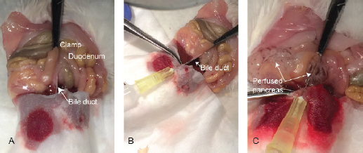
Figure 1. Courtesy: Bioprotocol 2016; 6(12) isolation and culture of the islets of Langerhans from rat pancreas.

[Click here to view]

Optimization of islet isolation and culture

Following are methods employed for the optimization of the isolation of rat pancreatic islets:

(A) Pancreas mincing and collagenase type IV digestion

(B) Pancreatic perfusion of collagenase type XI via the common bile duct (CBD) postduodenal occlusion

(C) Pancreas mincing and collagenase type XI digestion followed by cell straining and Ficoll gradient centrifugation.

(D) Pancreas mincing and collagenase type XI digestion followed by cell straining (500 μm), Ficoll gradient centrifugation, and cell straining (70 μm) (Graham et al., 2016; Samaddar et al., 2019).

Pancreas mincing and collagenase type IV digestion

Rat was sacrificed with the high dose of pentobarbital sodium I.P. The pancreas was isolated and then transferred to a sterile polypropylene Petri plate containing sterile Hank’s balanced salt solution (HBSS) with 20 mM of HEPES (N-2-hydroxyethylpiperazine-N-ethanesulfonic acid), 2 mM of CaCl2.2H2O, and penicillin–streptomycin–amphotericin B (100 IU/ml–100 μg/ml–2.5 μg/ml, respectively) solution. The superficial fatty tissues and blood clots were removed by HBSS wash and mincing of the pancreas followed by HBSS wash for three times. The minced tissue mass was transferred to a 50 ml of sterile conical-bottom centrifuge tube containing 5 ml of collagenase type IV (1 mg/ml; HiMedia, Mumbai) in Roswell Park Memorial Institute-1640 (RPMI 1640). The tube was incubated at 37°C in a water bath for 20 minutes with occasional shaking. After 20 minutes, the digestion was stopped by thrusting the tube in ice and adding 10% fetal bovine serum (FBS). The tube was centrifuged at 1,000 rpm for 10 minutes. The cell pellet was resuspended in warm RPMI with 10% FBS and seeded in a T25 culture flask (Nunc, Denmark). The islets were incubated at 37°C in 5% CO2 in a CO2 incubator (Forma, Thermo Scientific, Waltham, MA). Healthy islets (with smooth borders and no dark center) are handpicked using a 200-μl micropipette under the microscope and placed into a Petri plate containing fresh RPMI 1640 medium after 48 hours. These islets were counted and further evaluated for their functionality and viability.

Pancreatic perfusion of collagenase type XI via the CBD post duodenal occlusion

Rat was sacrificed with the high dose of pentobarbital sodium I.P., and an incision was made around the upper abdomen to expose the peritoneum. The liver was flipped over onto a tissue paper, and it was folded over to cover it. Using a curved forceps, the duodenum was located. The confluence, where the CBD enters the duodenum, was located and clamped with a hemostatic forceps to prevent the emptying of collagenase solution in the duodenum. Gently, 3 ml of ice-cold collagenase type XI (Sigma, St. Louis, MO) solution (1 mg/ml) in HBSS was injected through the CBD into the pancreas. The inflated pancreas was gently dissected and placed in a 50-ml conical centrifuge tube containing 2 ml of collagenase type XI solution. The tube was placed in a water bath at 37°C for 15 minutes and briefly shaken 2–3 times by hand during the incubation. After digestion, the tube was removed from the water bath and plunged immediately into ice to stop collagenase digestion, and 15 ml of fresh ice-cold HBSS was added to stop the digestion process completely. The HBSS was removed by centrifuging the tube at 1,000 rpm for 10 minutes, and then, the cell pellet was resuspended in warm RPMI with 10% FBS and seeded in T25 culture flask (Nunc, Denmark). The islets were incubated at 37°C in 5% CO2 in a CO2 incubator (Forma, Thermo Scientific, Waltham, MA). Healthy islets (with smooth borders and no dark center) are handpicked using a 200-μl micropipette under the microscope and placed into a Petri plate containing fresh RPMI 1640 medium after 48 hours. These islets were counted and further evaluated for their functionality and viability.

Pancreas mincing and collagenase type XI digestion followed by cell straining and Ficoll gradient centrifugation

Pancreatic digestion was carried similar to the previous method in a water bath with a slight modification in the digestion medium. Bovine serum albumin fraction V (Sigma, St. Louis, MO) was added to a final concentration of 2%. A 100 μl aliquot of the digestion mixture was withdrawn at a regular interval (5 minutes) and observed under the microscope to monitor the digestion process (Jacqueline et al., 2009). The complete detachment of exocrine tissues from the islets when observed indicates the completion of the digestion process. At this point, the digestion is stopped, and the mixture is centrifuged at 1,000 rpm for 10 minutes. The supernatant is removed, and the cell mass is resuspended in 10-ml warm RPMI. The digested pancreas is filtered through a sterile stainless steel mesh (500-μm pore size). The mesh is washed with 5–10 ml of additional cold RPMI to wash down any residual digested tissue leaving behind the undigested tissue. The tube is centrifuged to obtain the digested pancreas after removing the supernatant and resuspended gently in 10 ml of Histopaque® 1077 (Sigma, St. Louis, MO) at room temperature. This was then overlaid with 5 ml of RPMI 1640 at room temperature extremely gently forming a clear and sharp interface between the two liquids. The tube was centrifuged at 850 × g for 20 minutes with brakes off. It is just beneath the interface where the islets gather after centrifugation. The islets were gently removed from the gradient and transferred to a tube containing RPMI. The medium was removed by centrifugation to wash off the histopaque, and a fresh, warm complete RPMI 1640 containing 10% FBS was added to the islets. The islets were resuspended, seeded into a Petri plate, and incubated at 37°C in 5% CO2 for 48 hours followed by handpicking.

Pancreas mincing and collagenase type XI digestion followed by cell straining (500 μm), Ficoll gradient centrifugation, and cell straining (70 μm)

After density gradient centrifugation (in the previous step), the islets obtained were passed through a prewetted, inverted polypropylene 70-μm cell strainer (Cat. No. CLS431751; Corning, Corning, NY). The strainer was rewashed with fresh medium and turned upside down over a new petri dish containing 15 ml of fresh RPMI to rinse off the captured islets. This method eliminates the exocrine cells leaving behind islets only. The islets were incubated at 37°C in 5% CO2 for 48 hours and handpicked postincubation.

Assessment of islet viability and specificity

The islet viability was assessed by the trypan blue dye exclusion test, and the specificity of islets was determined by dithizone (DTZ) staining (Sigma, St. Louis, MO) (Samaddar et al., 2019). In the trypan blue dye exclusion assay, islets were exposed to the membrane-impermeant dye, trypan blue (0.1% w/v) for 15 minutes at 37°C. Dead and membrane-compromised cells took up the dye and appeared blue while healthy viable cells with intact membrane appeared colorless. For specificity, DTZ stock solution (39 mmol/l) was prepared by dissolving 100 mg of DTZ in 10 ml of DMSO, filtered, aliquoted, and stored at −20°C. Routine staining was carried out by adding 10 μl of DTZ stock to islets suspended in 1 ml of Krebs–Ringer bicarbonate (KRB) buffer (pH 7.4) with (4-(2-hydroxyethyl)-1-piperazineethanesulfonic acid) (HEPES) (10 mmol/l) and incubated at 37°C for 30 minutes.

Evaluation of islet protective potential assay

Glucose-stimulated insulin secretion assay (GSIS)

The isolated islets were cultured at 37°C in a humidified atmosphere of 5% CO2 in air in RPMI 1640 medium containing 11.1 mM of glucose, 10% of FBS, and antibiotics (50,000 IU/I penicillin and streptomycin) (Samaddar et al., 2019). The islets were seeded at a concentration of 50 islets per well in 12-well plates (Corning, Corning, NY), and the islets were washed thrice with KRB buffer and preincubated for 1 hour at 37°C. Grouping cells--- The islets were incubated for 1 hour at 37°C, and the aliquots of 10 μl were withdrawn from each well and assayed for insulin.

A total of 50 islets in triplicates (n = 3) were treated in the following manner:

Group 1: Glucose-free control – islets maintained in glucose-free KRB

Group 2: Normal glucose control – islets in 5.5 mM added glucose in KRB

Group 3: High glucose control – islets in 16.7 mM added glucose in KRB

Membrane integrity

After the islets were handpicked and incubated for 24 hours, they were exposed to the membrane-impermeant dye, trypan blue (0·1% w/v) for 15 minutes at 37°C. The presence of dye within cells was determined by light microscopy. Dead cells will take up the dye and appear blue owing to membrane damage, whereas the viable cells will remain unstained.

Effect of SMD on insulin secretion from cultured islets

The islets were cultured and treated with 5 mM of streptozotocin (Sigma, St. Louis, MO) in PBS, pH 7.4 with or without the SMD in low and high dose. Normal and high glucose-treated islets were also maintained in KRB and assayed for insulin.

A group of 50 islets each in triplicate (n = 3) were treated in the following manner:

Group 1: Normal control – islets maintained in glucose-free KRB, pH7.5

Group 2: Streptozocin (STZ) control – islets maintained in glucose-free KRB treated with 5 mM of STZ

Group 3: Normal glucose control – islets in 5.5 mM added glucose in KRB

Group 4: High glucose control – islets in 16.7 mM added glucose in KRB

Group 5: Saponin low dose – islets treated with SMD (10 μg/ml)

Group 6: Saponin high dose – islets treated with SMD (50 μg/ml)

Measurement of released and intra-islet insulin after treatment with STZ and SMD

A group of 50 islets each in triplicate (n = 3) were treated in the following manner:

Group 1: Normal control – islets maintained in glucose-free KRB, pH7.5

Group 2: STZ control – islets maintained in glucose-free KRB treated with 5 mM of STZ

Group 3: Saponin low dose – STZ control + SMD (10 μg/ml)

Group 4: Saponin high dose – STZ control + SMD (50 μg/ml)

Insulin was assayed by sandwich ELISA using a commercially available kit (Cat. No. 10-1250-01, Mercodia, Sweden).

Effect of the SMD on STZ-induced lipid peroxidation and nitric oxide (NO) formation

The assay mixture contained 0.5 ml of cell lysate, 1 ml of 0.5M KCl in 10 mM Tris-HCl, 0.5 ml of 30% trichloroacetic acid, and 0.5 ml of 52 mM thiobarbituric acid (TBA). The assay mixture was heated to 80°C for 30 minutes and after cooling to 0°C centrifuged at 800 x g for 10 minutes. The absorbance of the supernatant was measured at 532 nm. The levels of N-nitrosomethylaniline (NMA) were calculated using the following formula: (Absorbance at 532) – (Absorbance at 600) is Absorbance due to MDA-TBA abduct. The extinction coefficient of this MDA-TBA abduct at 532 nm is 155 mM−1cm−1. The concentration of Malondialdehyde (MDA) (mM) = (A532 – A600)/155

NO produced during STZ and saponin treatment was estimated spectrophotometrically as a formed nitrite (NO2). To measure the nitrite content, 100 μl of the cell lysate was incubated with 100 μl of Griess reagent (1% sulfanilamide in 0.1 mol/l HCl and 0.1% N-(1-naphthyl) ethylenediamine dihydrochloride) at room temperature for 10 minutes. Then, the absorbance was measured at 540 nm using a microplate reader. The nitrite content was calculated based on a standard curve constructed with NaNO2 (Ignarro et al., 1987).

Effect of SMD on high glucose-induced oxidative stress on islets

A group of 50 islets each in triplicate (n = 3) were treated in the following manner:

Group 1: Normal control – islets maintained in normal glucose (5.5 mM) medium

Group 2: High glucose control – islets maintained in high glucose (31 mM) medium

Group 3: Saponin low dose – STZ control + SMD (10 μg/ml)

Group 4: Saponin high dose – STZ control + SMD (50 μg/ml)

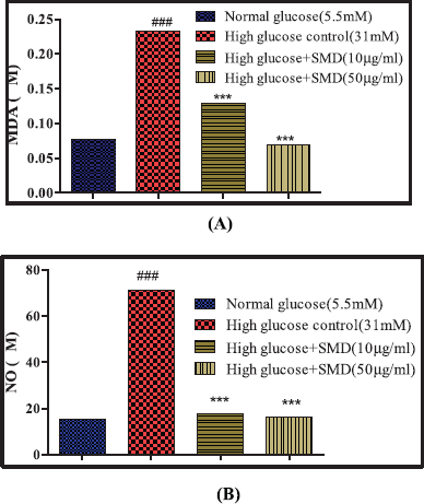
The islets were incubated for 24 hours at 37°C in 5% CO2. After incubation, the islets were lysed, as previous, and the lysate was used to estimate the formation of NO and MDA (lipid peroxidation).

Statistical Analysis

The results were expressed as mean ± SEM, and all the statistical comparisons were made by means of the one-way analysis of variance test followed by Tukey's multiple comparison tests (GraphPad Prism v5.0). The p-value < 0.05 was considered to indicate a statistically significant difference.
RESULTS

Saponin isolation and characterization

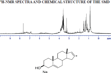
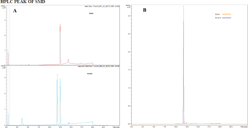
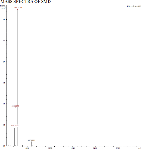
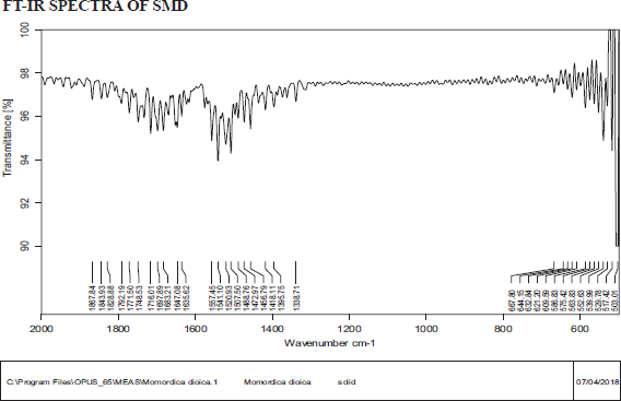
The saponin was isolated from the methanolic extract fruits of M. dioica, and the confirmation chemical test of M. dioica after isolated saponin was found to contain steroidal saponin (Fig. 2A, 2B, 3, 4 & 5).

Islet isolation and culture

After the optimized method for the isolation of rat pancreatic islets, the last method, which involves pancreas mincing and collagenase type XI digestion followed by cell straining (500 μm), Ficoll gradient centrifugation, and again cell straining (70 μm), produced the highest yield of purified islets with long-term viability (up to 3 days). The islets were completely digested and got released from the exocrine tissue. Cell straining avoided the entry of digested/undigested exocrine tissue into the islet pool, ensuring a very less damage to the islets. Through Ficoll gradient centrifugation, only islets could be selected which concentrate as a hazy band/ring immediately below the Ficoll–RPMI interface. Even though hand-picking of islets was carried out after 48 hours, almost 99% of exocrine cells were eliminated due to 70-μm straining because of which their detrimental effects on the islets were abolished. The islets obtained through this isolation technique were found to possess the maximum stability in terms of functionality and viability compared to the remaining three methods.

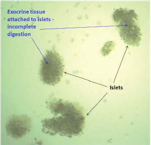
Other methods yielded lesser islets and poor stability and viability as they were incompletely digested (Fig. 6) and also got destroyed by the exocrine cells that found access into the islet pool as they were not specifically excluded from the digestion medium (straining and Ficoll gradient were not employed).

Figure 2. (A) and (B) indicates that the fraction collected contains a single component. Moreover, the saponin produced single peat at the retention time (Rt) of 11.5 minutes.

[Click here to view]

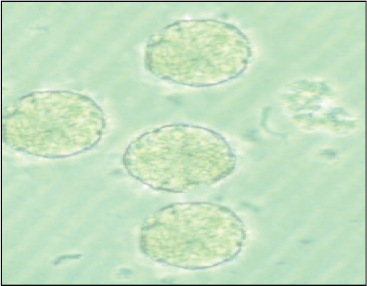
Healthy islets were recovered after 48-hour incubation with smooth rounded surface (Fig. 7) and were employed in further experiments. Larger islets are prone to develop hypoxic cells in their center, visibly distinguishable as darker cells compared to the surrounding tissue. Reducing the amount of media in the dish allowed increased oxygenation and reduced this effect (Table 1).

Assessment of islet viability and specificity

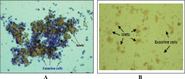
The islets were handpicked and incubated for 24 hours, and the islet viability and specificity were assessed by trypan blue dye exclusion test (0.1% w/v for 15 minutes at 37°C) and (DTZ, 10 μl) staining (Sigma, St. Louis, MO), respectively. The viable islets appear reddish-brown with DTZ, whereas dead exocrine cells stained blue with trypan blue (Fig. 8A and 8B).

Evaluation of islet protective potential assay

Glucose-stimulated insulin secretion assay

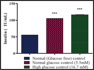
The isolated rat pancreatic islets were exposed in different concentrations of glucose, i.e., 5.5 and 16.7 mM. Insulin was assayed, and their response to glucose stimuli was noted (Fig. 9).

Values are expressed as mean ± SEM (n = 3). ***p < 0.05 Compared with the normal glucose-free control

Figure 3. Mass spectra of SMD: These fragments are compiled with the expected structure and found to be EI mass value at m/z M+: 293.

[Click here to view]

Figure 4. FT-IR spectra of SMD: Spectral data of FT-IR: 1,557.45 cm−1 (-OH stretching), 1,488 cm−1 (HC=CH stretching), 1,338.71 cm−1 (CH2-CH2 stretching), and 667.80 cm−1 (CH3 stretching).

[Click here to view]

Effect of SMD on insulin secretion from cultured islets

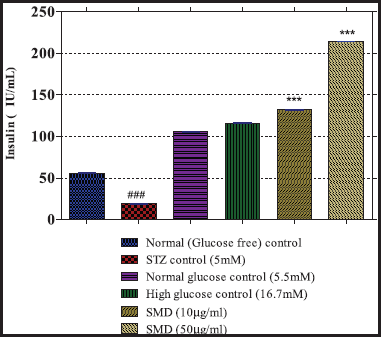
Insulin detection is one of the key indicators of β-cell existence and pancreatic β-cell protection by the SMD which was clearly seen to increase in a dose-dependent manner and data (Fig. 10).

Figure 5. 1H-NMR spectra and chemical structure of the SMD: The spectra exhibited aliphatic region, and it looks aliphatic cyclic organic compound fragment. Using the spectral data and chemical test, the basic skeleton of the structure of saponin was found to be steroidal saponin.

[Click here to view]

Figure 6. Incomplete digestion of islets illustrated by residual exocrine tissue attached to the islets exhibiting characteristic cloudy or hazy appearance over the islets.

[Click here to view]

Figure 7. Islets with uniform and entire boundary without central necrosis after 48 hours.

[Click here to view]

Table 1. Islet yield through different isolation methods.

[Click here to view]

Measurement of released and intra-islet insulin

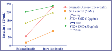
Glucose stimulates insulin secretion from islet β-cells but suppresses glucagon secretion from α-cell. A fine balance between insulin and glucagon secretion maintains the blood glucose levels within a narrow physiological range. In this assay, we looked at the released and intra-islet insulin to confirm the protective and regenerative potential of the drug on the pancreatic β-cells (Fig. 11).

Effect of SMD on STZ-induced lipid peroxidation and NO formation

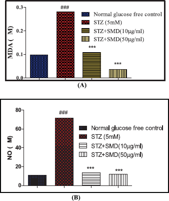
Nitric oxide can both promote and inhibit lipid peroxidation. By itself, NO acts as a potent inhibitor of the lipid peroxidation chain reaction by scavenging propagatory lipid peroxyl radicals. MDA levels were measured by the TBA method as previously reported by Konings and Drijver (1979). NO produced during STZ and saponin treatment was estimated spectrophotometrically as a formed nitrite (NO2) (Fig. 12A and 12B).

Effect of drug in high glucose-induced apoptosis on cultured islets

High glucose content can induce apoptosis and in the above experiment found that the SMD shows protection to the pancreatic islet as indicated by the measurement of MDA and NO levels (Fig. 13A and 13B).

Figure 8. (A) Islets stained with DTZ and trypan blue. (B) Islets in an environment of contaminating exocrine cells.

[Click here to view]

Figure 9. Glucose-stimulated insulin secretion assay.

[Click here to view]

Figure 10. Insulin secretions from cultured islets. Values are expressed as mean ± SEM (n = 3). ###p < 0.05 Compared with the normal control, ***p < 0.05 compared with the STZ control.

[Click here to view]

Figure 11. Comparison of the protective and regenerative potential of the drug on the pancreatic β-cells at SMD doses of 10 and 50 μg/ml. Values are expressed as mean ± SEM (n = 3). ###p < 0.05 Compared with the normal glucose-free control, ***p < 0.05 compared with the STZ control.

[Click here to view]

Figure 12. (A) Effect of SMD on STZ-induced LPO. Values are expressed as mean ± SEM (n=3). ###p < 0.05 Compared with the normal glucose-free control, ***p < 0.05 Compared with the STZ control. (B) Effect of SMD on STZ-induced NO. Values are expressed as mean ± SEM (n = 3). ###p < 0.05 Compared with the normal glucose-free control, ***p < 0.05 compared with the STZ control.

[Click here to view]

Figure 13. (A) Effect of SMD in high glucose-induced LPO on cultured islets. Values are expressed as mean ± SEM (n = 3). ###p < 0.05 Compared with the normal glucose group, ***p < 0.05 compared with the high glucose control group. (B) Effect of SMD in high glucose-induced NO on cultured islets. Values are expressed as mean ± SEM (n = 3). ###p < 0.05 Compared with the normal glucose group, ***p < 0.05 compared with the high glucose control group.

[Click here to view]


DISCUSSION

Through the ages, plants have always offered huge prospects toward the betterment of human health either by ameliorating disease conditions or enhancing normal physiological activity. Diabetes is one such metabolic disorder which is rampant throughout the world affecting individuals of every segment of the society. The complications of diabetes are multifactorial, which exacerbates the clinical conditions of the patients, eventually leading to death. Medicinal plants are extensively used as an alternative treatment strategy in the management and treatment of diabetes. It has been estimated that approximately 30% of diabetes worldwide have adopted the therapy offered by alternative and complementary medicine (Raman et al., 2012). In fact, the WHO has listed 21,000 medicinal plants, of which 150 are modestly used commercially (Joseph et al., 2011). The active constituents of herbal antidiabetic plants have been already reported to possess islet regeneration, insulin secretion, and overcoming resistance (Kavishankar et al., 2011).

The islets of Langerhans are the cluster of endocrine cells, and the target of immune-mediated destruction in type I diabetes has been reported (Thomas et al., 2016). The isolation of islets requires enzymatic and mechanical digestion of the exocrine tissue, and density gradient separation results in the isolation of 200–400 islets. Similarly, Sheng et al. (2009) reported a classical procedure that includes three steps: collagenase perfusion, pancreas digestion, and islet purification. The whole procedure takes 30–45 minutes for each individual, and a reasonable number of islets can be obtained in a relatively short period of time. Islet beta-cell replacement and regeneration are the keys approached for the treatment of diabetic patient. The current research has provided the proof of principle that the islet isolation was executed. The pancreas was removed and washed with HBSS, and the superficial fatty tissues and blood clots were removed. The most optimum method was found to be the pancreas mincing and collagenase type XI digestion followed by filtration gradient centrifugation and cell straining, and after density gradient centrifugation, the islets obtained were passed through a prewetted, inverted polypropylene 70-μm cell strainer. The islets were incubated in RPMI at 37°C in 5% CO2 for 48 hours, handpicked postincubation, and yielded the best islets both in terms of the quality and quantity.

A very well-known representative is Momordica charantia, belonging to the Cucurbitaceae family which is very popular as an antidiabetic plant. It is commonly known as bitter melon or bitter gourd whose fruit is very bitter on ripening. The extracts of M. charantia have been reported to possess hypoglycemic activity (Garau et al., 2003). The extracts of M. charantia and its various constituents have been reported to exert their hypoglycemic effect through various mechanisms such as utilization of glucose by peripheral and skeletal muscle (Uebanso et al., 2007), inhibition of glucose uptake from the intestine (Abdollah et al., 2010; Jeong et al., 2008), inhibition of the differentiation of adipocytes (Nerurkar et al., 2010), inhibition of primary gluconeogenic enzymes (Shibib et al., 1993), stimulation of important enzymes of HMP shunt pathway, and protection to the β-cells of the islets (Gadang et al., 2011).

Momordica dioica, which is a different species of the same genus and family, is abundant found in most of the states of India, Nepal, and the Himalayan region. It has been reported to possess type I and II antidiabetic activity and hypolipidemic activities, and many more were reported in the previous paper (Jha et al., 2017). These activities were investigated mainly focusing on in vivo models, but, in the present study, we investigated the antihyperglycemic activity in isolated rat pancreatic islets of Langerhans.

Islet viability and its function leading to failure use of antibiotics were examined (Bhonde et al., 2001). The viability and insulin production data showed that none of the antibiotics affect the viability and the function of the islets at their pharmacological concentrations. Free radical levels measured in terms of MDA, nitric oxide (NO), and reduced glutathione reveal that, except for a marginal increase in lipid peroxidation with tetracycline and slight increase in NO levels with streptomycin, none of these antibiotics affect the oxidative status of the cells. Similarly, cytokines play an important role in beta-cell failure (Yang et al., 2010). Cytokines, such as IL-1β, IFN-γ, TNF-α, leptin, resistin, adiponectin, and visfatin, have been shown to diversely regulate pancreatic β-cell function. NF-κB is a key signaling mechanism for pancreatic β-cell damage. Sulfuretin is one of the main flavonoids produced by Rhus verniciflua and reported to inhibit the inflammatory response by suppressing the NF-κB pathway. Rat insulinoma RINm5F cells and isolated rat islets were treated with IL-1β and IFN-γ to induce cytotoxicity reported (Song et al., 2010). Our intention was to explore the effectiveness and stability of the islet. Moreover, the islets obtained after handpicking were round with smooth and uniform boundary and without necrosis. They demonstrated decent specificity and viability through DTZ and trypan staining. The islets were also found to respond appropriately to GSIS assay, thereby qualifying for the functionality, specificity, and viability assessments. We have shown that the saponin has stimulated the secretion of insulin from islets significantly compared to STZ-treated islets. We further show the exposure of islets to STZ, and the saponin did not exhibit a reduction in insulin secretion which otherwise has happened with the STZ-treated islets. This is suggestive that beta-cell mass unaffected by STZ in the presence of the saponin. The results also revealed suppression in the formation of MDA, an index of Lipid peroxidation (LPO), and NO as opposed to the STZ-treated islets. We also assessed the effect of high glucose on isolated islets cultured in the presence of the saponin. It was reported that the high glucose-induced oxidative stress on islets leads to Reactive oxygen species (ROS) formation (Robertson et al., 2007), LPO (Turk et al., 1993), DNA damage (Wu et al., 2004), and apoptosis. In agreement to this, we revealed that the viability of islets in high glucose was reduced by almost two folds. However, the viability of islets treated with the saponin unaffected, as almost 70% islets were found to viable.

Finally, the results indicate that the saponin could be a promising sign and vindication of the antihyperglycemic activity of the SMD, thus proving our hypothesis.


CONCLUSION

To draw the conclusion of the research conducted, it can be commented based on the data generated; phytosaponin was isolated through Thin liquid chromatography (TLC) and characterized by HPLC, LCMS, FT-IR, and 1H-NMR. Using the different spectral analyses, the basic skeleton of the structure of saponin was found to be steroidal saponin, and further study can be done to elucidate the structure. The most optimum method was found to be the pancreas mincing and collagenase type XI digestion followed by cell straining, Ficoll gradient centrifugation, and cell straining. Moreover, the islet was confirmed by DTZ staining. GSIS showed that the islets secreted insulin in a dose-dependent manner with respect to the different concentrations of glucose compared with the normal control indicating its functionality. The SMD was found to stimulate insulin secretion and provide protection.


ACKNOWLEDGMENT

I would like to express heart full thanks to my beloved parents, guide for their support and blessings. And, I would, especially, like to thank who played an important role in this project: Dr. Vijay Kumar, Dr. Manoj Kumar, Mr. Joseph Vinod, and Miss. Ankita.


CONFLICT OF INTEREST

The authors declare that there is no conflict of interest related to this work.


FUNDING

None.


REFERENCES

Abdollah M, Zuki ABZ, Goh YM, Rezaeizadeh A, Noordin MM. The effects of Momordica charantia on the liver in streptozotocin induced diabetes in neonatal rats. Afr J Biotechnol, 2010; 9(31):5004–12.

Bhonde RR, Parab PB, Sheorin VS. An in vitro model for screening oral hypoglycemics. In vitro Cell Dev Biol Anim, 1999; 35(7):366–8. CrossRef

David G, Dolores, S. Greenspan's basic & clinical endocrinology. 9th edition. McGraw-Hill Medical, New York, NY, p 314, 2011; Chapter 17.

Dittrich HM, Dorsche HHV. The anatomical and histological investigation of the pancreas in the 19th century and pancreas research from discovery of islets. Anat Anz, 1978; 43(3):231–41.

Donath MY, Halban PA. Decreased beta-cell mass in diabetes: significance, mechanisms and therapeutic implications. Diabetologia, 2004; 47(3):581–9. CrossRef

Firdous, Haldar P, Bag AK. Evaluation of antidiabetic activity of saponin of Momordica dioica Roxb. Pharmacologyonline, 2009; 2:510–5.

Gadang V, Gilbert W, Hettiararchchy N, Horax R, Katwa L, Devareddy L. Dietary bitter melon seed increases peroxisome proliferators- activated receptor-γ gene expression in adipose tissue, down-regulates the nuclear factor- kB expression, and alleviates the symptoms associated with metabolic syndrome. J Med Food, 2011; 14:86–93. CrossRef

Garau C, Cummings E, Phoenix DA, Singh J. Beneficial effects and mechanism of action of Momordica charantia in the treatment of diabetes mellitus: a mini review. Int J Diabetes Metab, 2003; 11:46–55.

Graham K, Fynch S, Papas E, Tan C, Kay T, Thomas H: Isolation and Culture of the Islets of Langerhans from Mouse Pancreas. Bio-Protocol 2016; 6(12): e1840. DOI: 10.21769/BioProtoc.1840. CrossRef

Jacqueline OF. The isolation and purification of rodent pancreatic islets of Langerhans, Type 2 Diabetes. Methods Mol Biol, 2009; 560:37–42. CrossRef

Jeong J, Lee S, Hue J, Lee K, Nam SY, Yun YW. Effect of bitter melon (Momordica charantia) on antidiabetic activity in mice. Korean J Vet Res, 2008; 48(3):327–36.

Jha DK, Koneri R, Samaddar S. Antidiabetic activity of phytosaponin in STZ-Induced type I diabetes in rats. Res J Pharm Tech, 2019; 12(8):3919–26. CrossRef

Jha DK, Koneri R, Samaddar S. Potential Bio-Resources of Momordica dioica Roxb: a review. Int J Pharm Sci Rev Res, 2017; 45(2):203–9.

Joseph B, Jini D. Insight into the hypoglycaemic effect of traditional Indian herbs used in the treatment of diabetes. Res J Med Plant, 2011; 5(4):352–76. CrossRef

Kavishankar GB, Lakshmidevi N, Murthy SM, Prakash HS, Niranjana SR. Diabetes and medicinal plants-a review. Int J Pharm Biomed Sci, 2011; 2(3):65–80.

Konings AWT, Drijver EB. Radiation effects on membranes I: Vitamin E deficiency and lipid peroxidation. Radiat Res 1979; 80(3):494–501. CrossRef

Manisha M, Priyanjali D, Jayant L. Indian herbs and herbal drugs used for the treatment of diabetes. J Clin Biochem Nutr, 2007; 40:163–73. CrossRef

Nerurkar PV, Lee YK, Nerurkar VR. Momordica charantia (bitter melon) inhibits primary human adipocyte differenciation by modulation adipogenic genes. BMC Complement Altern Med, 2010; 10:34–9. CrossRef

Raman BV, Krishna NV, Rao NB, Saradhi PM, Rao BMV. Plants with antidiabetic activities and their medicinal values. Int Res J Pharm, 2012; 3(3):11–15.

Samaddar S, Koneri R, Varre JV, Jha DK. Triterpenoid saponin from Momordica tuberosa (Cucurbitaceae) stimulates insulin secretion from isolated mouse pancreatic islets and provides protection from streptozotocin and high-glucose induced injury. Ind J of Pharma Edu and Res, 2019; 53(3):511–20. CrossRef

Shibib BA, Khan A, Rahman R. Hypoglycaemic activity enzymes glucose-6-phosphates and fructose-1,6-bisphosphatase and elevation of both liver and red-cell shunt enzyme glucose-6-phospahte dehydrogenase. Biochem. J, 1993; 292:267–70. CrossRef

Singh RP, Padmavathi B, Rao AR. Modulatory influence of Adhatoda vesica (Justica adhatoda) leaf extract on the enzyme of xenobiotic metabolism, antioxidant status and lipid peroxidation in mice. Mol Cell Biochem, 2000; 213(1–2):99–109.

Uebanso T, Arai H, Taketani Y, Fukaya M, Yamamoto H, Mizuno A. Extracts of Momordica charantia suppress postprandial hyperglycemia in rats. J Nutr Sci Vitaminol, 2007; 53(6):482–86. CrossRef

Reference

Abdollah M, Zuki ABZ, Goh YM, Rezaeizadeh A, Noordin MM. The effects of Momordica charantia on the liver in streptozotocin induced diabetes in neonatal rats. Afr J Biotechnol, 2010; 9(31):5004-12.

Bhonde RR, Parab PB, Sheorin VS. An in vitro model for screening oral hypoglycemics. In vitro Cell Dev Biol Anim, 1999; 35(7):366-8.https://doi.org/10.1007/s11626-999-0108-3

David G, Dolores, S. Greenspan's basic & clinical endocrinology. 9th edition. McGraw-Hill Medical, New York, NY, p 314, 2011; Chapter 17.

Dittrich HM, Dorsche HHV. The anatomical and histological investigation of the pancreas in the 19th century and pancreas research from discovery of islets. Anat Anz, 1978; 43(3):231-41.

Donath MY, Halban PA. Decreased beta-cell mass in diabetes: significance, mechanisms and therapeutic implications. Diabetologia, 2004; 47(3):581-9.https://doi.org/10.1007/s00125-004-1336-4

Firdous, Haldar P, Bag AK. Evaluation of antidiabetic activity of saponin of Momordica dioica Roxb. Pharmacologyonline, 2009; 2:510-5.

Gadang V, Gilbert W, Hettiararchchy N, Horax R, Katwa L, Devareddy L. Dietary bitter melon seed increases peroxisome proliferators-activated receptor-γ gene expression in adipose tissue, down-regulates the nuclear factor- kB expression, and alleviates the symptoms associated with metabolic syndrome. J Med Food, 2011; 14:86-93.https://doi.org/10.1089/jmf.2010.0010

Garau C, Cummings E, Phoenix DA, Singh J. Beneficial effects and mechanism of action of Momordica charantia in the treatment of diabetes mellitus: a mini review. Int J Diabetes Metab, 2003; 11:46-55.

Graham K, Fynch S, Papas E, Tan C, Kay T, Thomas H: Isolation and Culture of the Islets of Langerhans from Mouse Pancreas. Bio-Protocol 2016; 6(12): e1840. DOI: 10.21769/BioProtoc.1840.https://doi.org/10.21769/BioProtoc.1840

Jacqueline OF. The isolation and purification of rodent pancreatic islets of Langerhans, Type 2 Diabetes. Methods Mol Biol, 2009; 560:37-42.https://doi.org/10.1007/978-1-59745-448-3_3

Jeong J, Lee S, Hue J, Lee K, Nam SY, Yun YW. Effect of bitter melon (Momordica charantia) on antidiabetic activity in mice. Korean J Vet Res, 2008; 48(3):327-36.

Jha DK, Koneri R, Samaddar S. Antidiabetic activity of phytosaponin in STZ-Induced type I diabetes in rats. Res J Pharm Tech, 2019; 12(8):3919-26.https://doi.org/10.5958/0974-360X.2019.00675.9

Jha DK, Koneri R, Samaddar S. Potential Bio-Resources of Momordica dioica Roxb: a review. Int J Pharm Sci Rev Res, 2017; 45(2):203-9.

Joseph B, Jini D. Insight into the hypoglycaemic effect of traditional Indian herbs used in the treatment of diabetes. Res J Med Plant, 2011; 5(4):352-76.https://doi.org/10.3923/rjmp.2011.352.376

Kavishankar GB, Lakshmidevi N, Murthy SM, Prakash HS, Niranjana SR. Diabetes and medicinal plants-a review. Int J Pharm Biomed Sci, 2011; 2(3):65-80.

Konings AWT, Drijver EB. Radiation effects on membranes I: Vitamin E deficiency and lipid peroxidation. Radiat Res 1979; 80(3):494- 501.https://doi.org/10.2307/3574991

Manisha M, Priyanjali D, Jayant L. Indian herbs and herbal drugs used for the treatment of diabetes. J Clin Biochem Nutr, 2007; 40:163-73.https://doi.org/10.3164/jcbn.40.163

Nerurkar PV, Lee YK, Nerurkar VR. Momordica charantia (bitter melon) inhibits primary human adipocyte differenciation by modulation adipogenic genes. BMC Complement Altern Med, 2010; 10:34-9.https://doi.org/10.1186/1472-6882-10-34

Raman BV, Krishna NV, Rao NB, Saradhi PM, Rao BMV. Plants with antidiabetic activities and their medicinal values. Int Res J Pharm, 2012; 3(3):11-15.

Samaddar S, Koneri R, Varre JV, Jha DK. Triterpenoid saponin from Momordica tuberosa (Cucurbitaceae) stimulates insulin secretion from isolated mouse pancreatic islets and provides protection from streptozotocin and high-glucose induced injury. Ind J of Pharma Edu and Res, 2019; 53(3):511-20.https://doi.org/10.5530/ijper.53.3.83

Shibib BA, Khan A, Rahman R. Hypoglycaemic activity enzymes glucose-6-phosphates and fructose-1,6-bisphosphatase and elevation of both liver and red-cell shunt enzyme glucose-6-phospahte dehydrogenase. Biochem. J, 1993; 292:267-70.https://doi.org/10.1042/bj2920267

Singh RP, Padmavathi B, Rao AR. Modulatory influence of Adhatoda vesica (Justica adhatoda) leaf extract on the enzyme of xenobiotic metabolism, antioxidant status and lipid peroxidation in mice. Mol Cell Biochem, 2000; 213(1-2):99-109.

Uebanso T, Arai H, Taketani Y, Fukaya M, Yamamoto H, Mizuno A. Extracts of Momordica charantia suppress postprandial hyperglycemia in rats. J Nutr Sci Vitaminol, 2007; 53(6):482-86.https://doi.org/10.3177/jnsv.53.482

Article Metrics
893 Views 67 Downloads 960 Total

Year

Month

Related Search

By author names